
1. Introduction
Newborn babies are very different from older children and adults in terms of how their body works, including their metabolism, immunity, and response to illness. Because of this, they need special care. Neonatal surgery refers to Surgeries performed during the first 28 days of life to treat conditions present at birth or those that develop soon after birth. These include congenital problems like oesophageal atresia, congenital diaphragmatic hernia, gastroschisis, omphalocele, intestinal atresia, anorectal malformations, and Hirschsprung disease, as well as emergency conditions such as necrotizing enterocolitis and spontaneous intestinal perforation [1–3].
The main idea behind neonatal surgery is that many birth defects that seem life-threatening at first and can actually be corrected with timely surgery, allowing the baby to survive and grow normally. Good outcomes depend on early diagnosis and proper treatment. Managing these babies requires a team approach involving neonatologists, paediatric surgeons, anesthesiologists, radiologists, geneticists, and specially trained nurses. Over time, improvements in prenatal diagnosis, neonatal intensive care, anesthesia, and surgical techniques have greatly improved survival rates [4–6].
However, neonatal surgery is still very challenging. It requires a deep understanding of how newborns function, careful surgical technique, and close monitoring after surgery. Another important issue is the difference in outcomes across the world. While survival has improved significantly in high-income countries, many babies in low- and middle-income countries still do not have access to proper surgical care. This gap highlights an important global health problem.
2. Historical Background
2.1 Ancient and Pre-Modern Observations
The understanding of congenital abnormalities goes back to very early times. Ancient Egyptian writings and Babylonian clay tablets describe babies born with visible defects, but these were often seen as signs or omens rather than medical problems that could be treated. Early thinkers like Aristotle and Galen described such abnormalities, but no attempts were made to treat them. In those times, babies born with severe defects were often abandoned or left to die, as this was accepted by society and law [1–3].
The first small steps toward treating these conditions started much later, around the 16th and 17th centuries. Fabricius ab Aquapendente in 1600 described a condition called imperforate anus and suggested that the blockage could be opened using a sharp instrument, although this idea was not widely practiced for many years. Later, in 1665, Frederik Ruysch described oesophageal atresia and recorded the first known case, but at that time there was still no treatment available. These early observations slowly laid the foundation for the development of neonatal surgery [4–6].
2.2 The 18th and 19th Centuries: Early Surgical Attempts
In the 18th century, a few early attempts were made to operate on newborn babies, although these were rare. One important example was in 1793, when Duret performed what is considered the first successful surgery for imperforate anus in an infant. He created a new opening for stool through a perineal approach. This was a major step forward because it showed, for the first time, that surgery in newborns was possible and that babies could survive after an operation [1–3].
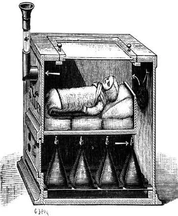
During the 19th century, interest in diseases of children slowly increased. This was partly due to the development of anesthesia by William T. G. Morton and James Young Simpson in the 1840s. However, using anesthesia in newborns was still very risky, so most surgeons avoided operating on them. In 1888, Harald Hirschsprung described a condition of severe bowel enlargement in infants, now known as Hirschsprung disease. At that time, there was no effective treatment, and affected infants usually did not survive [4–6].
Efforts to treat conditions like oesophageal atresia during this period were unsuccessful, and all such cases resulted in death. In fact, more than 95% of newborns with intestinal obstruction died. This high mortality was mainly due to the lack of safe anesthesia, poor surgical techniques, absence of intravenous fluids, and no proper postoperative care facilities such as intensive care units [7– 9].
2.3 The 20th Century: The Birth of Modern Neonatal Surgery
Modern neonatal surgery really began in the early part of the 20th century. Key Pioneers William E. Ladd – established pediatric surgery as a specialty Recognize as the father of paediatric surgery in North America. Robert Gross a student of Ladd performed the first successful ligation of PDA in 1938. C. Everett Koop – contributed significantly to neonatal surgical care. One of the most important developments was safer anesthesia for children, introduced by pioneers like William E. Cotton and Cameron Haight. A major breakthrough came in 1939, when Cameron Haight successfully operated on a newborn with oesophageal atresia and tracheo-oesophageal fistula at the University of Michigan. This was the first time such a complex condition was treated successfully, and it proved that newborn babies with serious defects could survive after surgery. This marked the beginning of modern neonatal surgery [1–3].
In the years that followed, many important surgical advances were made. In 1948, Orvar Swenson and Bill introduced the pull-through operation for Hirschsprung disease, which became the standard treatment. Surgeons like Willis J. Potts improved techniques for joining the intestine (anastomosis) in newborns. At the same time, William E. Ladd and Robert E. Gross at Boston Children’s Hospital developed systematic approaches to treat many neonatal conditions. Their work helped establish paediatric surgery as a separate and specialized field [4–6]. Peter Paul Rickham, & J. Herbert Johnston published the ‘Bible of Neonatal Surgery’ in 1969 detailing breakthroughs from the Alder Hey Children’s Hospital in Liverpool
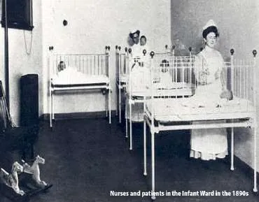
Paediatric surgery became an official subspecialty during this period. Important organizations like the American Academy of Pediatrics Section on Surgery and the British Association of Paediatric Surgeons were formed to promote education and research in this area. Over time, formal training programs, dedicated paediatric surgical units, and specialized neonatal intensive care units (NICUs) were developed during the 1950s and 1960s. These advances greatly improved the care and survival of newborns requiring surgery [7–9].

William E. Ladd

. C. Everett Koop

Robert E. Gross
2.4 Late 20th Century Advances
In the later part of the 20th century, there were major improvements in neonatal surgical care. One of the most important advances was the introduction of total parenteral nutrition (TPN) by Stanley Dudrick in 1968. This allowed babies who could not take feeds, especially after major intestinal surgery, to receive proper nutrition through intravenous routes and survive. At the same time, improvements in neonatal intensive care—such as mechanical ventilation, surfactant therapy, extracorporeal membrane oxygenation (ECMO), and high-frequency ventilation—greatly increased survival in premature babies and those with severe breathing problems. Because of these advances, more sick newborns could safely undergo surgery [1–3].
Another important change was in neonatal anaesthesia. Earlier, it was wrongly believed that newborns did not feel pain, and many surgeries were performed with inadequate pain control. This changed after the landmark study by K. J. S. Anand and P. R. Hickey in 1987, which showed that proper anaesthesia reduces stress and improves outcomes in neonates. This led to better pain management and safer perioperative care for newborns [4–6].
During the 1980s and 1990s, minimally invasive surgery also began to develop in neonates. After the success of laparoscopic surgery in adults, paediatric surgeons started using these techniques in infants. Karl Georgeson and his team showed that procedures like laparoscopic Nissen fundoplication and laparoscopic surgery for Hirschsprung disease could be done safely in children. This marked the beginning of minimally invasive neonatal surgery, which continues to grow and improve today. [7–9]
2.5 Foetal Surgery: The Frontier of Prenatal Intervention
One of the most remarkable developments in modern neonatal surgery is foetal surgery, where treatment is given to the baby even before birth while the mother is still pregnant. This idea was once thought to be impossible, but it became a reality in 1981 when Michael R. Harrison and his team at the University of California, San Francisco performed the first successful open foetal surgery for a condition called obstructive uropathy. After this, foetal surgery was gradually used for other serious conditions such as congenital diaphragmatic hernia (CDH), myelomeningocele, sacrococcygeal teratoma, and twin-to-twin transfusion syndrome [1–3].
A major breakthrough came with the Management of Myelomeningocele Study (MOMS) published in 2011. This study showed that repairing myelomeningocele before birth led to better neurological outcomes compared to surgery done after birth. This provided strong scientific evidence supporting the role of foetal surgery in selected cases [4–6].
In recent years, newer and less invasive techniques have been developed. These include procedures like fetoscopic tracheal occlusion for CDH and other percutaneous (through the skin) foetal treatments. These advances have expanded the number of conditions that can be treated before birth and represent an important extension of neonatal surgical care into the prenatal period [7–9].
3. Scope of Neonatal Surgery
3.1 Classification of Neonatal Surgical Conditions
Neonatal surgical conditions may be usefully classified according to their anatomical system, their embryological basis, or their urgency of intervention. From an urgency perspective, conditions may be categorized as true surgical emergencies requiring intervention within hours of birth (oesophageal atresia with respiratory compromise, tension pneumothorax from CDH, gastroschisis with bowel compromise), urgent conditions requiring surgery within 24 to 48 hours (intestinal atresias, imperforate anus without fistula, large omphalocele), and conditions that may be managed on a semi-elective basis within the first weeks of life (smaller abdominal wall defects, uncomplicated Hirschsprung disease).
3.2 Major Neonatal Surgical Conditions
Oesophageal Atresia and Tracheo-Oesophageal Fistula
Oesophageal atresia (OA), with or without tracheo-oesophageal fistula (TOF), is a condition seen in about 1 in 3,000 to 4,500 newborn babies. In this condition, the food pipe (oesophagus) does not form properly. The most common type is Gross type C, where the upper part of the oesophagus ends blindly and the lower part is connected to the windpipe (trachea). This type accounts for about 85% of all cases [1–3].
Babies with this condition usually present soon after birth with excessive salivation, choking, coughing, and bluish discoloration (cyanosis), especially during feeding. Doctors may also find that a nasogastric tube cannot be passed into the stomach, which helps in diagnosis. Many of these babies also have other associated anomalies, commonly grouped under the VACTERL association. These associated problems are seen in up to 50% of cases and play an important role in deciding treatment and outcome [4–6].Treatment is surgical. The main aim is to connect the two ends of the esophagus and close the abnormal connection with the trachea.
Congenital Diaphragmatic Hernia (CDH)
Congenital diaphragmatic hernia (CDH) occurs in about 1 in 2,000–4,000 newborns and still has a significant mortality of around 20–30%, even in well-equipped centres. It happens because the diaphragm does not form properly during early pregnancy, allowing abdominal organs like the intestine and stomach to move into the chest. This affects lung development, leading to small lungs (pulmonary hypoplasia) and high pressure in lung blood vessels (pulmonary hypertension). Nowadays, treatment focuses first on stabilising the baby with gentle ventilation, medications, and sometimes Extracorporeal Membrane Oxygenation before surgery is done. Once stable, the defect is repaired either through an open abdominal approach or minimally invasive (thoracoscopic) surgery [1–3].
Abdominal Wall Defects
The two main abdominal wall defects in newborns are gastroschisis and omphalocele.
Gastroschisis occurs when there is a hole beside the umbilicus, and the intestines come out without any covering. It occurs in about 1 in 3,000 births and is seen more often in younger mothers. Treatment involves protecting the exposed bowel, gradually placing it back into the abdomen (sometimes using a silo), and then closing the defect.
Omphalocele, on the other hand, occurs at the umbilicus, and the herniated organs are covered by a sac. These babies often have other associated problems, especially heart defects, seen in up to 30% of cases. Because of this, management is more complex and needs careful planning [4–6].
Intestinal Obstruction
Common causes of intestinal blockage in newborns include intestinal atresia, malrotation with volvulus, meconium ileus, and Hirschsprung disease. Duodenal atresia occurs in about 1 in 5,000 births and is often associated with Down syndrome (trisomy 21). It is treated by connecting the two ends of the intestine (duodenoduodenostomy).
Jejunoileal atresia usually occurs due to reduced blood supply to the intestine before birth and is treated by removing the affected part and joining the healthy ends. Malrotation with volvulus is a surgical emergency because the intestine can twist and lose blood supply. It is treated with Ladd's procedure, which untwists the bowel and prevents further complications [7–9].
Anorectal Malformations (ARM)
Anorectal malformations include a range of defects where the anus and rectum do not develop properly. These can vary from simple conditions, which can be corrected with minor surgery, to more complex ones involving abnormal connections with the urinary or genital tract. The Krickenbeck classification is used to classify these conditions. The standard surgery for most cases is posterior sagittal anorectoplasty (PSARP), developed by Alberto Peña and de Vries [10–12].
Necrotising Enterocolitis (NEC)
Necrotising enterocolitis (NEC) is the most common serious intestinal emergency in newborn intensive care units, especially in premature babies. It has a mortality of about 20–30%, and even higher in severe cases. The disease involves damage to the intestine due to poor blood supply, infection, and inflammation. Surgery is needed if there is a hole in the intestine, worsening condition despite treatment, or severe disease. This usually involves removing the damaged bowel and creating a temporary stoma. In very small or unstable babies, a drain may be placed as a temporary measure [13–15].
4. Epidemiology and Global Burden of Neonatal Surgical Disease
4.1 Global Incidence and Prevalence
Congenital anomalies (birth defects) are an important cause of death in newborns worldwide. According to the World Health Organization, around 303,000 babies die every year within the first month of life due to these conditions. This accounts for nearly 11% of all neonatal deaths. Many of these conditions can actually be treated with surgery if proper care is available. Overall, major congenital anomalies occur in about 2–3% of all live births globally, although not all of them need surgery immediately after birth [1–3].
Some important neonatal surgical conditions have known incidence rates. Oesophageal atresia occurs in about 1 in 3,500 births. Congenital diaphragmatic hernia occurs in about 1 in 2,500– 4,000 births. Gastroschisis has been increasing in recent years and is now seen in about 1 in 2,000– 5,000 births. Intestinal atresias occur in about 1 in 5,000 births. Hirschsprung disease occurs in about 1 in 5,000 births and is more common in boys. Anorectal malformations occur in about 1 in 2,500–5,000 births. necrotising enterocolitis affects about 7–13% of very low birth weight babies (less than 1,500 g) [4–6].
4.2 Burden of Disease in Low- and Middle-Income Countries (LMICs)
The burden of neonatal surgical diseases is much higher in low- and middle-income countries (LMICs). These countries have higher birth rates but limited access to prenatal diagnosis, neonatal intensive care, and surgery. About 94% of deaths due to congenital anomalies occur in these regions. In many places, conditions that are easily treated in developed countries can lead to death because proper facilities are not available. It is estimated that around 1.7 million children under five die every year from conditions that could be treated with surgery [7–9].
The Lancet Commission on Global Surgery highlighted that about five billion people worldwide do not have access to safe and affordable surgical care. Studies from regions like sub-Saharan Africa show that many hospitals lack even basic equipment. For example, fewer than 40% of centres have pulse oximeters, and less than 20% have trained paediatric surgeons. As a result, death rates for conditions like intestinal obstruction and abdominal wall defects can be more than 50%. This clearly shows the gap between the number of patients and the available healthcare resources [10–12].
4.3 Economic and Social Impact
Neonatal surgical diseases not only cause death but also lead to long-term health problems and financial burden. Babies who survive conditions like congenital diaphragmatic hernia, oesophageal atresia, or necrotising enterocolitis may face long hospital stays, feeding problems, breathing issues, and developmental delays. Treatment is expensive, especially in high-income countries. For example, in the United States, the cost of treating gastroschisis can range from $50,000 to $75,000, and in complicated cases it can go above $250,000 [13–15].
In LMICs, families often have to pay for treatment themselves, which can push them into poverty or make them avoid seeking care altogether. When measured using disability-adjusted life years (DALYs), neonatal surgical conditions have a very high impact because they affect newborns who have their whole life ahead. Studies have shown that improving access to neonatal and paediatric surgery is cost-effective and can give benefits similar to vaccination programs in improving public health [16–18].
5.1 Cardiovascular Physiology
At birth, a newborn baby’s circulation changes a lot. Before birth, the baby’s blood flow is different, with high pressure in the lungs and special pathways like the ductus arteriosus and foramen ovale that allow blood to bypass the lungs. After birth, when the baby starts breathing, the pressure in the lungs decreases and normal circulation begins. However, in the first few days, lung pressure can still remain high and may increase again due to problems like low oxygen, acidosis, cold stress, or pain. This can lead to a condition called Persistent Pulmonary Hypertension of the Newborn, where blood flows abnormally and causes low oxygen levels [1–3].
The newborn heart also works differently. Babies have a higher heart rate (about 120–160 beats per minute), but their heart muscle is less flexible and cannot increase output easily. This means cardiac output mainly depends on heart rate. If the heart rate drops (bradycardia), the cardiac output also drops quickly. Procedures like suctioning or handling the airway can trigger this. The neonatal heart is also more sensitive to drugs, especially anaesthetic agents, because it has less reserve compared to adults [4–6].
5.2 Respiratory Physiology
Newborn babies have a higher risk of breathing problems, especially during and after surgery. Their chest wall is soft and flexible, which makes it harder to maintain proper lung expansion. Their diaphragm also gets tired easily because it is not fully developed. The amount of air left in the lungs after breathing out (functional residual capacity) is low, which can lead to airway collapse and poor oxygen exchange [7–9].
Newborns mainly breathe through their nose, so any blockage like secretions or a feeding tube can cause breathing difficulty. They also need more oxygen than adults (about double per kg), but their oxygen reserves are low, so they can become hypoxic quickly. Apnoea (temporary stopping of breathing) is common, especially in premature babies and after anaesthesia, so close monitoring is very important after surgery [10–12].
5.3 Thermoregulation
Newborn babies lose heat very easily. This is because they have a large body surface area, very little fat, and cannot shiver effectively, especially if they are premature. Instead, they produce heat using brown fat (brown adipose tissue), but this is limited and can get exhausted quickly. Heat loss can occur through the environment, contact with cold surfaces, air movement, and evaporation [13–15].If a baby becomes cold during surgery (hypothermia), it can lead to problems like poor blood clotting, increased oxygen demand, slow drug metabolism, weak immunity, and delayed recovery. Therefore, it is very important to keep the baby warm during surgery. This is done by warming the operation room, using warming blankets, warming IV fluids, covering the baby
5.4 Fluid and Electrolyte Balance
Newborn babies have a higher amount of water in their body compared to adults. In fact, total body water is around 75–80% of body weight at birth, compared to about 60% in adults. A larger portion of this is outside the cells (extracellular fluid), which gradually decreases in the first week of life as the baby passes more urine. However, the kidneys of newborns are not fully developed, so they cannot concentrate or dilute urine properly. Their kidney filtration rate (GFR) is also much lower at birth and improves slowly over time [1–3].Because of this, giving fluids to newborns needs careful calculation and monitoring. Too much fluid can cause problems like lung swelling (pulmonary oedema) and heart-related issues, while too little fluid can lead to dehydration, kidney injury, and low blood sugar. Usually, fluids are started at about 60–80 mL/kg/day on the first day and gradually increased to around 120–150 mL/kg/day by day 3–4, depending on the baby’s condition [4–6].
5.5 Metabolic and Glucose Balance
Newborns, especially premature or low birth weight babies, have low energy reserves and are at risk of low blood sugar (hypoglycaemia). Their bodies cannot produce enough glucose quickly, so blood sugar needs to be maintained above safe levels. For this reason, glucose-containing fluids are routinely given, and blood sugar is checked regularly. On the other hand, high blood sugar (hyperglycaemia) can also occur, especially in sick premature babies receiving intravenous nutrition, and this can lead to complications like infections and brain bleeding [7–9].Other electrolyte problems are also common. Calcium levels may drop, especially after blood transfusion. Problems like low sodium (hyponatraemia), high potassium (hyperkalaemia), and acidosis can occur and need correction. Newborns have limited ability to balance acids and bases, so acidosis can develop quickly during stress, blood loss, or poor circulation [10–12].
5.6 Haematological and Coagulation Characteristics
Newborn babies mainly have foetal haemoglobin (HbF), which holds on to oxygen more tightly than adult haemoglobin. This is useful before birth but after birth it can make oxygen delivery to tissues slightly less efficient. Normal haemoglobin levels are relatively high at birth but decrease over the next few weeks, leading to what is called physiological anaemia of infancy [13–15].
Clotting factors in newborns are also lower than in adults, especially vitamin K–dependent factors. This increases the risk of bleeding, which is why vitamin K is routinely given at birth. Platelet counts are usually normal in healthy babies but can be low in premature or sick newborns. Because of these factors, surgeons need to carefully monitor bleeding and transfusion needs during surgery [16–18].
5.7 Pharmacokinetics and Drug Handling
Drugs behave differently in newborns compared to older children and adults. The liver enzymes that break down drugs are not fully developed, and kidney function is also immature, so drugs are cleared more slowly. Newborns also have more body water, so water-soluble drugs spread more widely in the body. In addition, protein levels in the blood are lower, which affects how drugs are carried in the bloodstream [19–21].
Because of all these differences, medicines stay longer in the body and can have stronger effects. Therefore, all drugs in newborns must be given carefully based on weight and age, with close monitoring to avoid side effects [22–24].
6. Principles of Neonatal Surgical Care
6.1 Prenatal Diagnosis and Counselling
Modern neonatal surgical care often starts even before the baby is born. During pregnancy, routine ultrasound scans—especially in the second trimester—can detect many birth defects early. This helps doctors counsel the parents, plan the delivery, and prepare the surgical and neonatal care team in advance. In more complex cases, additional imaging like foetal MRI is used to get better details, especially for conditions such as congenital diaphragmatic hernia or large tumours. Genetic counselling is also important when there is a risk of inherited or chromosomal disorders. Ideally, delivery should take place in a centre where immediate newborn surgical care is available, or the baby should be transferred there as soon as possible .
6.2 Resuscitation and Stabilisation
Before any surgery, the baby must be stabilised. The first step is to ensure proper breathing, good oxygen levels, and stable blood circulation. Problems like low body temperature, low blood sugar, electrolyte imbalance, infection, and acidosis should be corrected before surgery, as they can increase complications. A tube is usually passed into the stomach (nasogastric tube) to remove air and prevent vomiting or aspiration. Intravenous access is established, often through an umbilical venous catheter in newborns, to give fluids and medicines. If infection is suspected, blood tests are done and antibiotics are started early.
6.3 Anaesthetic Considerations
Anaesthesia in newborns requires special care and expertise. A breathing tube is inserted gently using the correct size to avoid injury. Anaesthetic drugs are given in lower doses because newborns are more sensitive to their effects, especially on the heart. Pain relief is very important, and medicines like opioids are used carefully to avoid breathing problems. Regional anaesthesia techniques, such as caudal or spinal blocks, can also be used to provide good pain relief and reduce the need for stronger drugs. Muscle relaxants may be used when needed for surgery. Throughout the procedure, the baby’s temperature is closely monitored and maintained using warming methods [7–9].
6.4 Intraoperative Principles
During surgery in newborns, very gentle handling of tissues is extremely important because their tissues are delicate. Surgeons take great care to control bleeding and avoid unnecessary handling of organs. Since the operating area is very small, fine instruments and sometimes magnifying lenses or microscopes are used for better precision. Very fine sutures (like 5-0 to 7-0) are used to reduce tissue injury. Whenever suitable, minimally invasive techniques like laparoscopy or thoracoscopy are preferred because they cause less stress to the baby, give better cosmetic results, and allow faster recovery.
It is also important to keep the duration of surgery as short as possible, as long procedures can lead to problems like hypothermia, low blood sugar, and breathing instability. In some very sick babies, surgeons may not perform the full surgery immediately. Instead, they do a temporary procedure to stabilise the baby and plan the final surgery later when the baby is stronger. This approach, called damage control surgery, is useful in conditions like intestinal perforation, necrotising enterocolitis, and complex abdominal wall defects.
6.5 Postoperative Care
Care after surgery is just as important as the operation itself and is usually done in a neonatal intensive care unit (NICU). Many babies need breathing support with a ventilator after major surgery, and this is gradually reduced as the baby improves. Pain relief is given using a combination of medicines to keep the baby comfortable. Doctors closely monitor fluids, electrolytes, blood sugar, and blood counts during this period [7–9.Feeding is started slowly once the baby’s intestine begins to function again. Until then, nutrition is given through intravenous methods like total parenteral nutrition (TPN). Doctors also watch carefully for complications such as leakage from surgical joins, infections, lung problems like pneumothorax, or recurrence of fistula. Early detection and management of these complications are very important [10–12].
6.6 Long-Term Follow-Up
Babies who undergo major neonatal surgery need long-term follow-up. This includes monitoring growth, nutrition, and overall development. Some children may have ongoing digestive problems such as reflux or bowel movement issues, especially in conditions like Hirschsprung disease or after complex surgeries. Breathing problems and developmental delays may also occur in some cases [13–15].
Regular follow-up helps in early detection and management of these issues. As these children grow older, their care needs to be smoothly transferred from paediatric to adult healthcare services. This transition is important to ensure continued management of long-term complications and to maintain a good quality of life [16–18].
7. Challenges in Developing Countries
7.1 Infrastructure and Resource Constraints
Providing neonatal surgical care in low- and middle-income countries (LMICs) is very difficult due to lack of proper facilities and resources. One of the biggest problems is the shortage of neonatal intensive care units (NICUs). After surgery, newborns need close monitoring and support, but in many hospitals this is not available. Even if the surgery is done well, babies may still die due to problems like infection, breathing difficulty, low body temperature, or low blood sugar. Studies from regions like sub-Saharan Africa show that many hospitals do not even have dedicated neonatal beds, and basic monitoring equipment such as pulse oximeters may not be available consistently .
7.2 Workforce Shortages
Another major challenge is the lack of trained doctors. There are very few paediatric surgeons in LMICs. According to the World Federation of Associations of Pediatric Surgeons, some regions like sub-Saharan Africa have only about 0.1 paediatric surgeons per million people, compared to 7–10 per million in developed countries like the UK and USA. There is also a shortage of trained paediatric anaesthesiologists, which makes neonatal surgery even more difficult. Training opportunities are limited, and many doctors move to higher-income countries for better opportunities, leading to a “brain drain” and worsening the shortage .
7.3 Delayed Presentation and Referral
In many developing countries, babies with surgical problems are often brought to hospitals very late. By the time they reach proper care, their condition may have worsened significantly. This delay happens due to many reasons such as long distance from hospitals, poor transport facilities, lack of awareness among parents, cultural beliefs, financial problems, and delays within the healthcare system itself. Conditions like intestinal obstruction or volvulus become more serious with delay, leading to damaged bowel and higher risk during surgery. This makes treatment more difficult and reduces the chances of survival
7.4 Financial Barriers
Neonatal surgery is very expensive, especially in low- and middle-income countries (LMICs). The cost includes surgery, NICU stay, investigations, medicines, blood products, and long hospital admission. In many countries, there is little or no health insurance to cover these expenses. Because of this, families often have to pay from their own pocket. This can be a huge burden, and sometimes families have to choose between treatment and basic needs like food and housing. In many cases, access to life-saving surgery depends more on money than on medical need .
7.5 Anesthetic Challenges
Providing safe anaesthesia for newborns is also difficult in LMICs. Many hospitals lack modern equipment and have problems like unreliable electricity and limited drug supply. Older machines and drugs like halothane are still commonly used instead of newer, safer options. Monitoring devices such as pulse oximeters and capnography may not always be available, which increases the risk during surgery. In some places, anaesthesia is given by non-specialists or staff with limited training, leading to higher complication rates [4–6].
7.6 Sepsis and Infection
Infections are a major cause of death after neonatal surgery in LMICs. This is mainly due to poor infection control practices, lack of proper antibiotics, and the presence of resistant bacteria in hospitals. Surgical complications like wound infections, leakage from bowel connections, and abdominal infections are more common in such settings. Overcrowding, poor sterilisation, and lack of proper equipment also contribute. Additionally, babies may already be at higher risk due to maternal infections like HIV, syphilis, or malaria [7–9].
7.7 Documentation and Data
Another important problem is the lack of proper records and data. Many hospitals do not maintain detailed records of neonatal surgeries, and follow-up of patients is often incomplete. Without good data, it becomes difficult to understand how many children are affected, how well treatments are working, and what improvements are needed. This also makes it harder to plan better healthcare policies. Setting up proper data systems, such as hospital databases or national registries, is essential to improve neonatal surgical care in these regions [10–12].
8. Future Perspectives
8.1 Technological Innovation
The future of neonatal surgery is expected to improve a lot with new technologies. Smaller and more precise robotic surgical systems are being developed, which may help surgeons perform delicate operations in tiny babies with better accuracy. New techniques like 3D bioprinting may allow the creation of customised materials, such as meshes for abdominal wall repair or even tissue substitutes for organs like the oesophagus, designed specifically for each patient [1–3].Imaging during surgery is also becoming more advanced. Techniques like near-infrared fluorescence imaging using dyes such as Indocyanine Green help surgeons see blood flow in tissues in real time. This can help in deciding which part of the bowel is healthy before joining it, reducing complications. Other innovations like high-resolution ultrasound during surgery, augmented reality, and artificial intelligence are also being explored to make surgery safer and more effective [4–6].
8.2 Genetic and Molecular Advances
Advances in genetics are helping doctors understand the causes of many congenital conditions better. Technologies like whole genome sequencing can identify genetic changes responsible for diseases such as congenital diaphragmatic hernia, oesophageal atresia, and Hirschsprung disease. This allows earlier diagnosis during pregnancy, better counselling for families, and identification of associated syndromes [7–9].In the future, these advances may also lead to targeted treatments, including gene therapy, where the underlying genetic problem is corrected. Although still in early stages, this area has great potential for changing how congenital diseases are treated [10–12].
8.3 Foetal and Neonatal Medicine Integration
The line between foetal and neonatal care is becoming less clear as treatments are now possible even before birth. Techniques like Foetal Endoluminal Tracheal Occlusion are being used for severe congenital diaphragmatic hernia, and ongoing studies like the TOTAL trial are evaluating their benefits. These procedures aim to improve lung development before the baby is born [13– 15].Other areas of research include stem cell therapy to improve organ development and the development of artificial placenta systems, which could support extremely premature babies outside the womb. These innovations may completely change how we manage high-risk pregnancies and neonatal conditions in the future [16–18].
8.4 Global Surgery Initiatives
There is a big difference in access to neonatal surgical care between high-income countries and low- and middle-income countries (LMICs). To reduce this gap, efforts are needed at many levels, including training, funding, and government support. The Lancet Commission on Global Surgery has recommended that everyone should have access to safe and affordable surgery by 2030. Organisations like World Federation of Associations of Pediatric Surgeons, Global Initiative for Children's Surgery, and Smile Train are helping by setting up training centres and improving surgical care in developing regions [1–3].
In places where specialists are not available, task-sharing is being used. This means general surgeons or trained healthcare workers are taught to perform basic but life-saving procedures like colostomy or drainage for necrotising enterocolitis. Technology is also helping through telemedicine, online teaching, and remote mentoring. Partnerships between hospitals in developed and developing countries are helping to improve skills and infrastructure over time [4–6].
8.5 Quality Improvement and Outcome Research
In the future, more focus will be placed on improving the quality of care and measuring outcomes. Large research networks like Canadian Pediatric Surgery Network, Pediatric Surgical Research Collaborative, and ERNICA are collecting data from many centres. This helps doctors understand which treatments work best, identify complications early, and improve overall care [7–9]. There is also increasing importance given to patient-reported outcomes, such as quality of life after surgery. These help doctors understand the long-term impact of treatment, not just survival [10– 12].
8.6 Regenerative Medicine and Organoids
Regenerative medicine is an exciting future area in neonatal surgery. Scientists are working on growing tissues in the lab using stem cells. For example, intestinal organoids (mini versions of the intestine grown in the lab) may help treat conditions like short bowel syndrome in the future. Early experiments in animals have shown promising results [13–15].
There have also been attempts to create replacement structures like the trachea using tissue engineering. Research is ongoing to develop artificial oesophagus, heart tissue patches, and even lung models for conditions like congenital diaphragmatic hernia. Although still in early stages, these advances may completely change how congenital conditions are treated in the coming decades [16–18].
9. Conclusion
Neonatal surgery is one of the greatest achievements in modern medicine. In the past, many babies born with serious congenital problems could not survive, but today, most of these conditions can be treated successfully with good outcomes and acceptable quality of life. This progress has happened over many years, starting from early milestones like the first successful oesophageal atresia repair by Cameron Haight in 1939, to the present era of advanced techniques such as minimally invasive surgery, robotic surgery, foetal interventions, and regenerative medicine. These improvements have been made possible by better understanding of newborn physiology, safer anaesthesia, improved surgical techniques, and advances in neonatal intensive care [1–3].
However, many challenges still remain. Around the world, babies in low- and middle-income countries (LMICs) continue to face higher risk because of limited access to proper surgical care. There is a large gap between what can be achieved in well-equipped hospitals and what is available in resource-poor settings. Addressing this gap is not only a medical issue but also an important ethical and public health concern. It requires better infrastructure, more trained specialists, improved financial support systems, and proper data collection to guide future improvements [4– 6].
References
| 1. | Langer JC. Neonatal surgery. In: Coran AG, Adzick NS, Krummel TM, Laberge JM, Shamberger RC, Caldamone AA, editors. Pediatric Surgery. 7th ed. Philadelphia: Elsevier Saunders; 2012. p. 1–18. |
|---|---|
| 2. | Haight C, Towsley H. Congenital atresia of the esophagus with tracheoesophageal fistula: extrapleural ligation of fistula and end-to-end anastomosis of esophageal segments. Surg Gynecol Obstet. 1943;76:672–88. |
| 3. | Swenson O, Bill AH Jr. Resection of rectum and rectosigmoid with preservation of the sphincter for benign spastic lesions producing megacolon: an experimental study. Surgery. 1948;24(2):212– 20. |
| 4. | Gross RE, Ladd WE. Abdominal Surgery of Infancy and Childhood. Philadelphia: W.B. Saunders; 1941. |
| 5. | Anand KJS, Hickey PR. Pain and its effects in the human neonate and fetus. N Engl J Med. 1987;317(21):1321–9. |
| 6. | Dudrick SJ, Wilmore DW, Vars HM, Rhoads JE. Long-term total parenteral nutrition with growth, development, and positive nitrogen balance. Surgery. 1968;64(1):134–42. |
| 7. | Adzick NS, Thom EA, Spong CY, Brock JW 3rd, Burrows PK, Johnson MP, et al. A randomized trial of prenatal versus postnatal repair of myelomeningocele. N Engl J Med. 2011;364(11):993– 1004. |
| 8. | Harrison MR, Golbus MS, Filly RA, Callen PW, Katz M, de Lorimier AA, et al. Fetal surgery for congenital hydronephrosis. N Engl J Med. 1982;306(10):591–3. |
| 9. | Georgeson KE, Fuenfer MM, Hardin WD. Primary laparoscopic pull-through for Hirschsprung's disease in infants and children. J Pediatr Surg. 1995;30(7):1017–21. |
| 10. | Metkus AP, Filly RA, Stringer MD, Harrison MR, Adzick NS. Sonographic predictors of survival in fetal diaphragmatic hernia. J Pediatr Surg. 1996;31(1):148–51. |
| 11. | World Health Organization. Congenital anomalies [Internet]. Geneva: WHO; 2023 [cited 2024 Nov 10]. Available from: https://www.who.int/news-room/fact-sheets/detail/congenital-anomalies. |
| 12. | Bickler SW, Weiser TG, Kassebaum N, Higashi H, Chang DC, Barendregt JJ, et al. Global burden of surgical conditions. In: Debas HT, Donkor P, Gawande A, Jamison DT, Kruk ME, Mock CN, editors. Essential Surgery: Disease Control Priorities. 3rd ed. Washington: World Bank; 2015. p. 19–40. |
| 13. | Meara JG, Leather AJ, Hagander L, Alkire BC, Alonso N, Ameh EA, et al. Global Surgery 2030: evidence and solutions for achieving health, welfare, and economic development. Lancet. 2015;386(9993):569–624. |
| 14. | Ameh EA, Bickler SW, Lakhoo K, Nwomeh BC, Poenaru D, editors. Paediatric Surgery: A Comprehensive Text for Africa. Seattle: Global HELP Organisation; 2011. |
| 15. | Poenaru D. Getting the job done: analysis of the impact and coverage of the Smile Train program in alleviating the global burden of cleft disease. World J Surg. 2013;37(7):1562–70. |
| 16. | de Vries PA, Pena A. Posterior sagittal anorectoplasty. J Pediatr Surg. 1982;17(5):638–43. |
| 17. | Engum SA, Grosfeld JL. Intestinal atresia and stenosis. In: Grosfeld JL, O'Neill JA, Fonkalsrud EW, Coran AG, editors. Pediatric Surgery. 6th ed. Philadelphia: Mosby; 2006. p. 1269–88. |
| 18. | Neu J, Walker WA. Necrotizing enterocolitis. N Engl J Med. 2011;364(3):255–64. |
| 19. | Stoll BJ, Hansen NI, Bell EF, Shankaran S, Laptook AR, Walsh MC, et al. Neonatal outcomes of extremely preterm infants from the NICHD Neonatal Research Network. Pediatrics. 2010;126(3):443–56. |
| 20. | Walker GM, Laing IA, Ogston SA, Bhatt P, Morison G, Clark J, et al. The epidemiology of gastroschisis. Arch Dis Child Fetal Neonatal Ed. 2006;91(5):F326. |
| 21. | Spitz L. Esophageal atresia. Lessons I have learned in a 40-year experience. J Pediatr Surg. 2006;41(10):1635–40. |
| 22. | Deprest J, Gratacos E, Nicolaides KH; FETO Task Group. Fetoscopic tracheal occlusion (FETO) for severe diaphragmatic hernia: evolution of a technique and preliminary results. Ultrasound Obstet Gynecol. 2004;24(2):121–6. |
| 23. | Snoek KG, Reiss IK, Greenough A, Capolupo I, Urlesberger B, Wessel L, et al. Standardized postnatal management of infants with congenital diaphragmatic hernia in Europe: The CDH EURO Consortium consensus – 2015 update. Neonatology. 2016;110(1):66–74. |
| 24. | Laberge JM, Flageole H, Nguyen LT, Guttman FM. The VATER association: effects of tracheoesophageal fistula repair and reflux control in children. J Pediatr Surg. 1996;31(3):460–2. |
| 25. | Hirschsprung H. Stuhlträgheit Neugeborener in Folge von Dilatation und Hypertrophie des Colons. Jahrb Kinderheilkd. 1888;27:1–7. |
| 26. | Global Initiative for Children's Surgery. Global Initiative for Children's Surgery optimal resources for children's surgical care: a manual for program implementation. J Pediatr Surg. 2019;54(2):128– 37. |
| 27. | Krickenbeck International Classification Group. International classification of anorectal malformations. J Pediatr Surg. 2005;40(8):1391. |
| 28. | Agrawal N, Pratap A, Tiwari A, Sharma S. Outcome of neonatal surgery in developing countries. J Pediatr Surg. 2011;46(2):246–50. |
| 29. | Ozgediz D, Langer M, Kisa P, Poenaru D. Pediatric surgery as an essential component of global health. Semin Pediatr Surg. 2016;25(1):3–9. |
| 30. | Holcomb GW 3rd, Ostlie DJ, editors. Ashcraft's Pediatric Surgery. 7th ed. Philadelphia: Elsevier; 2020. |
| 31. | Bjornsson S, Gunnarsdottir A, Gesundheit B, Palsson G. Nationwide study of esophageal atresia in Iceland, 1990–2006: a population-based study. J Pediatr Surg. 2007;42(1):166–9. |
| 32. | Pena A, Hong AR. Advances in the management of anorectal malformations. Am J Surg. 2000;180(5):370–6. |
| 33. | Zamakhshary M, To T, Guan J, Langer JC. Risk of incarceration of inguinal hernia among infants and young children awaiting elective surgery. CMAJ. 2008;179(10):1001–5. |
| 34. | Heij HA. Current status and future prospects of neonatal surgery. Semin Pediatr Surg. 2014;23(5):237–40. |
| 35. | Antonoff MB, Saltzman DA, Acton RD, Ferreira LM, Nath S, Olney CM, et al. Obtaining surgical consent for neonatal repair of esophageal atresia: does gestational age affect the decision? J Pediatr Surg. 2009;44(10):1857–64. |