
Abstract
Neonatal cholestasis is a serious clinical disease characterized by conjugated hyperbilirubinemia, which indicates significant hepatobiliary dysfunction and necessitates rapid examination. This chapter provides a complete description of newborn cholestasis, focusing on the most urgent and prevalent cause, biliary atresia. We look at pathophysiology, various etiologies, clinical presentation, diagnostic options, and therapeutic strategies for these interrelated disorders. Early identification, especially of biliary atresia, is critical because immediate surgical intervention (Kasai hepatic portoenterostomy) is required for native liver survival. This chapter also examines the long-term prognosis and the critical function of liver transplantation in the treatment of end-stage liver disease caused by these disorders.
Neonatal Cholestasis
Neonatal cholestasis is characterized by persistent yellowish discoloration of sclera, skin and pale colored stool with dark urine. It is a clinical and biochemical condition defined as impaired bile flow, leading to an accumulation of conjugated (direct) bilirubin in the blood. Biochemically, it is defined as a conjugated (direct) bilirubin level that exceeds 1.0 mg/dL (17 mmol/L) or more than 20% of the total bilirubin level. Physiologically, bile acids, bilirubin, and other chemicals are retained in the liver and accumulate in the systemic circulation because of neonatal cholestasis, which is a failure of bile production or flow. It must be differentiated from benign physiological jaundice because it is a pathological condition. It is a sign of severe underlying liver or biliary disease, never a normal physiological condition. This condition presents a diagnostic and therapeutic challenge due to its wide spectrum of underlying etiologies, ranging from surgically correctable conditions like biliary atresia to complex metabolic and genetic disorders. A delay in diagnosis and treatment can lead to irreversible liver damage, liver failure, and even death.
Neonatal cholestasis affects around one out of every 2,500 live newborns. It is a true pediatric emergency because the time for appropriate treatment of the most serious causes is limited. The key problem for the treating clinicians is to quickly distinguish between medical (intrahepatic) and surgical (extrahepatic) reasons. Infants presenting with pale stools, yellow discoloration of urine, and predominantly conjugated hyperbilirubinemia (direct/conjugated bilirubin levels exceeding 1.0 mg/dL when total serum bilirubin (TSB) is 5.0 mg/dL, or more than 20% of TSB if TSB is above 5.0 mg/dL) should be further evaluated for potential obstructive cholangiopathy.
Pathophysiology
Bile flow is a multifaceted complex process. Hepatocytes, cholangiocytes (epithelial cells of bile ducts), and a network of transporters are all involved in the intricate process of bile flow. Defects at any stage of this system might result in cholestasis. Hepatocytes are poisoned by the retained bile components, which lead to fibrosis, oxidative stress, inflammation, and ultimately cirrhosis. There are serious clinical repercussions when the intestines lack bile led to malabsorption or inadequate digestion and absorption of dietary fats and fat-soluble vitamins (A,D,E,K). There may be steatorrhea, failure to thrive, and certain vitamin deficiency diseases (such as coagulopathy from vitamin K insufficiency). Jaundice and other systemic issues are brought on by elevated circulating bilirubin. In severe circumstances, elevated bile acids may contribute to multi-organ dysfunction and produce pruritus, or itching.
Etiology and Differential Diagnosis
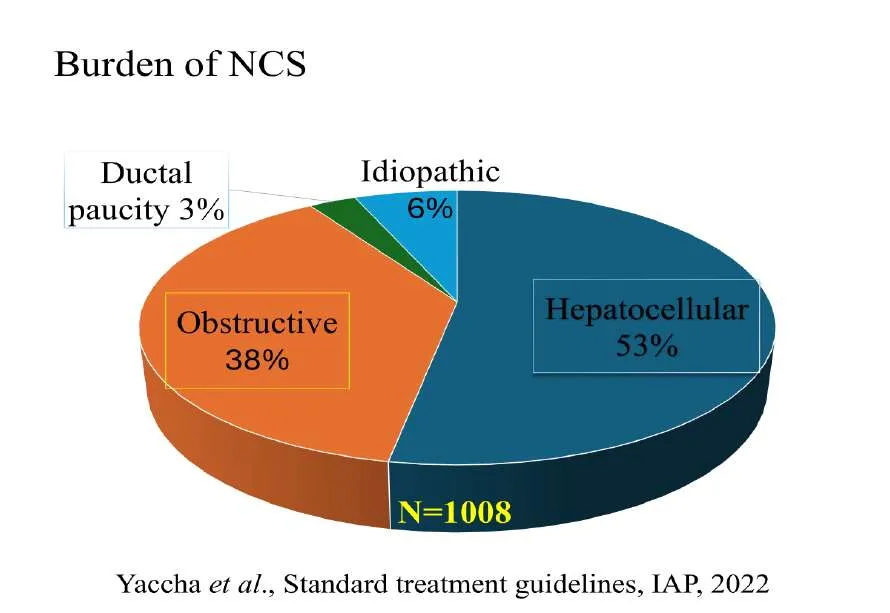
Neonatal cholestasis (NCS) has a wide range of etiology. With 25–40% of patients, biliary atresia is the most common surgical cause of neonatal cholestasis. Burden of NCS described by Yachha, et al depicted in Picture-1. The causes of neonatal cholestasis are often described as a "Pandora's Box" due to their immense variety. In general, the causes of cholestasis can be divided into two categories: Extrahepatic causes, which are primarily surgical; and Intrahepatic causes, which are primarily medical.
The several medical and surgical causes of newborn cholestasis are listed in Table-1 below, along with a differential diagnosis.
| Extrahepatic (Surgical) Biliary Atresia: | Intrahepatic (Medical) Infections: |
|---|---|
| A progressive inflammatory disease leading to complete obstruction of the extrahepatic bile ducts | Bacterial (sepsis, urinary tract infections) Viral (cytomegalovirus, herpes simplex virus, parvovirus B19) Protozoal (Toxoplasma) |
| Choledochal Cyst: | Metabolic Disorders: |
| Congenital dilations of the bile ducts which can cause obstruction. These typically present with jaundice or an abdominal mass and are surgically correctable | Inborn errors of metabolism, including alpha-1-antitrypsin deficiency, galactosemia, tyrosinemia, Niemann-Pick disease, and bile acid synthesis defects, etc. |
| Spontaneous perforation of bile ducts | Genetic/Hereditary Disorders: Alagille syndrome Progressive Familial Intrahepatic Cholestasis (PFIC) Benign recurrent intrahepatic cholestasis (BRIC) Cystic fibrosis, Ductal plate malformations Down syndrome, etc. |
Neonatal Sclerosing Cholangitis Parenteral Nutrition-Associated Cholestasis (PNAC):
Complication in premature and critically ill infants receiving prolonged total parenteral nutrition. Bile duct stenosis / Cholelithiasis / Idiopathic Neonatal Hepatitis: a diagnosis of exclusion.
Inspissated bile / Mucus plug / Extrinsic
compression.
Clinical Presentation and Red Flags
Persistent jaundice is the characteristic of newborn cholestasis. Acholic (pale, grey, or white) feces are a major warning sign because they strongly indicate a total or almost complete biliary obstruction, most frequently biliary atresia. Other important signs that should be evaluated right once include low weight gain (failure to thrive), an enlarged liver (hepatomegaly), and dark, tea-coloured urine. Important diagnostic hints include a history of maternal disease, preterm, or symptoms in other organ systems (e.g., respiratory problems in cystic fibrosis, cardiac problems in Alagille syndrome).
Diagnostic Workup and Evaluation
The diagnosis procedure needs to be organized and quick. When an infant's jaundice lasts longer than 14–21 days, the first step is always to fractionate bilirubin levels. Following the confirmation of cholestasis, a methodical assessment is conducted to determine the precise cause.
Laboratory Investigations: A wide range of blood tests are carried out, such as liver function tests (AST, ALT, GGT), coagulation profile (INR/PT), metabolic screening (for tyrosinemia, galactosemia, and alfa-1-antitrypsin deficiency), TORCH (Toxoplasma, Other, Rubella, Cytomegalovirus, Herpes) screening, and possibly bile acid analysis.
Imaging: When looking for symptoms of biliary atresia such as the "triangular cord sign," gallbladder enlargement, and choledochal cysts, abdominal ultrasonography is usually the first imaging modality used. After five days of phenobarbital/UDCA priming, hepatobiliary scintigraphy (HIDA scan) can be used to measure bile excretion into the intestines, however its precision is not perfect.
Liver Biopsy: A liver biopsy is an essential diagnostic procedure that frequently distinguishes biliary atresia from intrahepatic causes (such as newborn hepatitis) with high accuracy. Intraoperative Cholangiography (IOC): During exploratory surgery, IOC is used to definitively diagnose biliary atresia. The Kasai portoenterostomy operation is conducted right away if it is determined that the bile ducts are atretic.
Management and Treatment Strategies
Neonatal cholestasis management entails both comprehensive supportive care and targeted therapy of the underlying cause. Surgical Management: Surgical correction is necessary for biliary atresia and choledochal cysts, including mechanical obstruction or compression.
Medical Management: Treatment for intrahepatic causes focuses on controlling the specific ailment, such as antibiotics for infections, enzyme replacement therapy for Wolman disease, or dietary restrictions for galactosemia. Ursodeoxycholic acid (UDCA) is a drug that is frequently used to protect liver cells and enhance bile flow in the majority of cholestatic disorders. Supportive Care: Aggressive nutritional support, such as high-calorie formulas and supplements of fat-soluble vitamins (A, D, E, and K) to prevent malabsorption and failure to thrive, is a key component of care for all types of newborn cholestasis.
Liver Transplantation: Liver transplantation is a life-saving operation for illnesses including severe alfa-1-ATD and many forms of PFIC, and it is the only treatment for instances that advance to end-stage liver disease or if specific medication is unavailable or inadequate (e.g., failed Kasai surgery).
Prognosis and Long-Term Outcomes of Neonatal Cholestasis
Neonatal cholestasis has a very erratic outcome. While biliary atresia patients frequently have a difficult journey involving the Kasai procedure, long-term complications like cholangitis and portal hypertension, and a high likelihood of requiring a liver transplant later in childhood or adulthood, infants with conditions like idiopathic neonatal hepatitis typically recover well. For all aetiologies, early diagnosis and referral to expert pediatric hepatology centres enhance results. Early detection of biliary atresia and increased native liver survival rates have been demonstrated by public health initiatives, such as the use of infant stool colour cards in parental education.
Summarize, newborn cholestasis is a serious pediatric illness that needs to be treated quickly. A quick, methodical diagnostic approach is required due to its many causes. In order to prevent irreparable liver damage and guarantee the greatest long-term health results for affected newborns, prompt diagnosis and targeted treatment are crucial, especially for time-sensitive disorders like biliary atresia.
Biliary Atresia
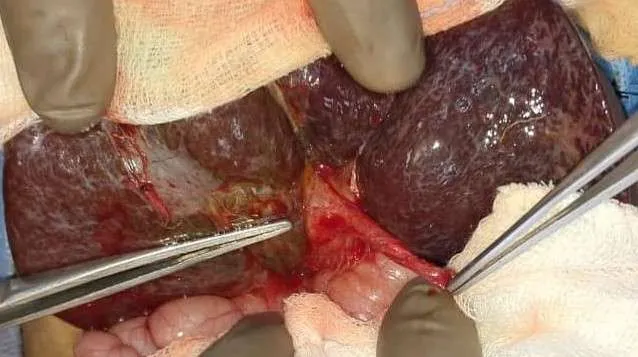
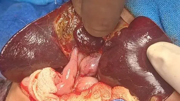
Professor John Burns originally described biliary atresia in 1817. Up to 1960, these patients had a bad prognosis. Professor Morio Kasai's 1950 procedure marked a significant development in the surgical treatment of biliary atresia. Biliary atresia is a progressive obstructive cholangiopathy of unknown cause that affects both the intrahepatic and extrahepatic bile ducts (panductular illness). It is the most severe and time-sensitive cause of neonatal cholestasis, defined by a distinct, unusual, progressive fibroinflammatory illness of the extrahepatic bile ducts that leads to their full obliteration shortly after delivery [Picture-2]. It is the most common and serious cause of newborn cholestasis, an obstructive cholangiopathy. Its origin is mainly unknown, but evidence suggests a mix of genetic predispositions, putative viral triggers, and immune-mediated ductal damage that occurs in utero or soon after birth. Approximately 1 in 6,000 to 18,000 live babies have biliary atresia. 25–40% of all cases of newborn cholestasis are caused by it. Asians and African Americans are more likely to have it, with a small female predominance (1.4:1 to 1.7:1). 11–20% of instances of EHBA are linked to congenital abnormalities, and 7.5% of cases had polysplenia. Biliary atresia with splenic malformations (BASM) is the syndromic category that includes cases of both biliary atresia and splenic abnormalities.
Etiopathogenesis
Although the precise origin of biliary atresia is unknown, several factors, including genetic, immunological, developmental, infectious, and environmental ones, are believed to be involved.
Occult Viral Infections: Due to viral-induced neoantigens on the biliary epithelium, occult viral infections with viruses such reovirus 3, cytomegalovirus (CMV), rotavirus C, human papillomavirus, and retroviruses may cause an immunological reaction.
Developmental Failure- Failure in the biliary ducts' recanalization process during development is known as developmental failure.
Ductal Plate Malformation Theory- Biliary ducts with inadequate support may burst, resulting in severe fibrosis that leads to ductal obliteration.
Exposure to Environmental Toxins- The illness may be exacerbated by exposure to environmental toxins.
Classification of Biliary Atresia
Devenport M. et al. divided biliary atresia into four major clinical groups based on etiopathogenesis:
1. Syndromic Biliary Atresia (Embryonic Type): Congenital problems such polysplenia, intestinal malrotation, situs inversus, preduodenal portal vein, interrupted inferior vena cava, and heart malformations are linked to this kind. A poor prognosis is linked to Biliary Atresia Splenic Malformation Syndrome (BASM syndrome), which accounts for 10–20% of cases. It develops while the foetus is still developing.
2. Cystic EHBA: This kind, which occurs in 5–10% of cases, typically arises during pregnancy and involves cystic dilatation of the biliary tree.
3. Cytomegalovirus-Associated Biliary Atresia Cytomegalovirus infection, which has either a prenatal or postnatal beginning and is characterized by increased IgM anti-CMV antibodies, is associated with about 10% of cases.
4. Isolated (Non-syndromic) Biliary Atresia With a prenatal or postnatal onset, this is the most prevalent form, accounting for 70–80% of cases.
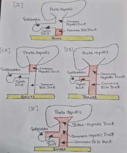
Depending on the location of extrahepatic bile duct obstruction, the Japanese Biliary Atresia Society (JBAS) divided biliary atresia into three fundamental kinds. [Picture-3]
Type I: Atresia of common bile duct (approx. 5%), proximal duct is intact.
Type II: Atresia of hepatic duct (approx. 2%).
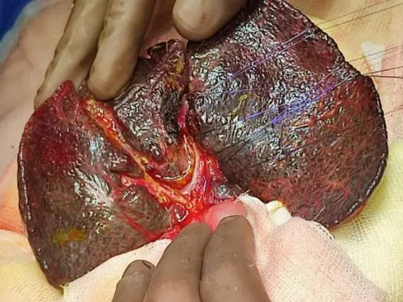
Type IIa - Obliteration of the common hepatic duct with intact common bile duct. Type IIb - Obliteration of the cystic duct, common bile duct and common hepatic duct. Type III: Atresia of bile duct at the porta hepatis (commonest type >90%). In this type, the cystic duct, the common bile duct, the hepatic ducts, and intrahepatic ducts are obliterated. Grossly, the liver is usually hard, swollen, and green or dark in colour during the initial stages of biliary atresia. The gallbladder may be completely atretic or appear tiny and packed with white fluid. The liver gets hard and nodular as the illness progresses. There are unique microscopic features that help differentiate biliary atresia from other forms of newborn hepatitis.
Clinical Presentation
Biliary atresia babies usually look healthy at birth and generally eat well at first. These are the main clinical indicators:
Persistent Jaundice and conjugated hyperbilirubinemia Jaundice worsens and does not resolve by 2-3 weeks of age. Measure total and conjugated bilirubin levels in any infant who has jaundice for more than two weeks. Acholic Stools Pale, gray, or white faces indicate a complete lack of bile pigments in the gut. This is a critical red flag. Parental teaching with stool colour cards has proven beneficial in countries such as Taiwan for early detection.
Dark Urine: Because conjugated bilirubin is excreted.
Hepatomegaly: Usually, a firm, enlarged liver can be felt. Delayed diagnosis might lead to advanced liver disease symptoms such as ascites and splenomegaly.
Diagnosis of Biliary Atresia
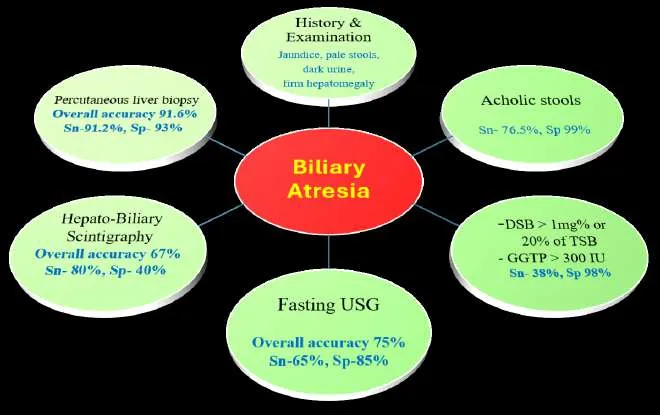
The diagnosis process must be quick. Once conjugated hyperbilirubinemia has been proven, specific procedures are taken. (Picture -4) depicts the outline of the diagnostic work-up. Laboratory Tests: A complete hemogram, coagulation profile (PT/INR), liver function tests (LFT), viral serology for hepatitis A, B, and C, serological tests for TORCH infections, and some additional metabolic disease tests (Alpha-1 Antitrypsin Level), thyroid function tests, genetic studies, and urine examination for sugar reduction may be required. Typically, high AST, ALT, and, most importantly, an elevated gamma-glutamyl transferase (GGT) level, which frequently distinguishes BA from other intrahepatic causes such as PFIC type 1 and 2, where GGT may be normal or low.
Radiological Imaging:
Abdominal Ultrasound: First-line imaging modality for assessing jaundiced babies. It is used to examine the presence and features of the gallbladder, detect intrahepatic or extrahepatic bile duct dilatation, and determine liver parenchymal echogenicity. Key findings suggestive of biliary atresia are:
Triangular Cord Sign: Atretic bile ducts in the porta hepatis.
GB Ghost Triad: Abnormal gallbladder size, wall thickening, and mucosal irregularities. Hepatic Artery hypertrophy and resistive index: An increased diameter of the hepatic artery is another indicative of biliary atresia.
Observation of postprandial gallbladder contraction may exclude the diagnosis of biliary atresia.
In advanced cases, ultrasound may reveal signs of portal hypertension.
Magnetic Resonance Cholangio-Pancreatography (MRCP)- Less commonly employed in clinical practice, it has lately emerged as a reliable diagnostic technique for biliary atresia in certain individuals, offering detailed vision of the biliary tree and accompanying anomalies.
Hepatic Scintigraphy with Hepatobiliary Imino-di-acetic Acid (HIDA) or Di-isopropyl
Imino-di-acetic Acid (DISIDA)- The presence of a radiotracer in the gastrointestinal system rules out biliary atresia, but the absence of a radiotracer is just suggestive of it. Patients should receive oral phenobarbital (5 mg/kg) for 5 days before to imaging to increase the test's sensitivity.
Duodenal Aspiration for Bile- Not regularly performed but can be considered in specialized situations. Aspirating the contents of the duodenum to detect the presence of yellow bilirubin pigment. If bilirubin is discovered, the diagnosis of biliary atresia is eliminated.
Endoscopic Retrograde Cholangio-Pancreatography (ERCP)- rarely used but can be considered in specific situation.
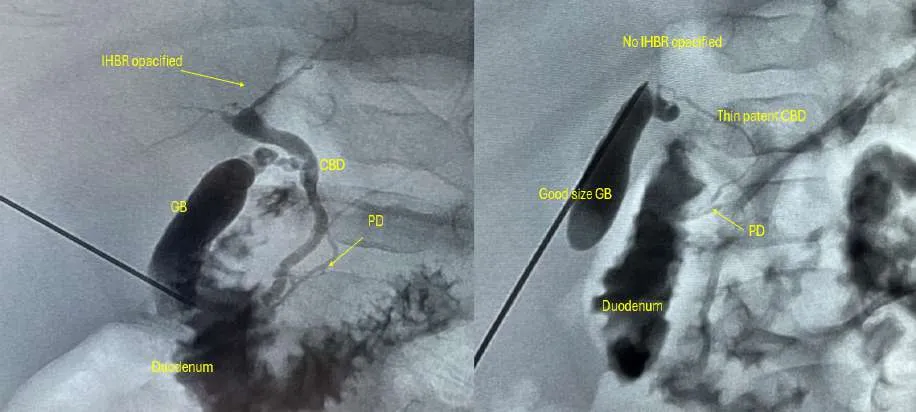
Percutaneous Transhepatic Cholecysto-Cholangiography (PTCC)*- Under sedation, a USG-guided gall bladder puncture is performed, and contrast is introduced into a remnant bile duct to examine the biliary tree. Mostly utilized in questionable cases where decent size gall bladder is present (its routine practice at author`s institute) [Picture-5].
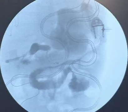
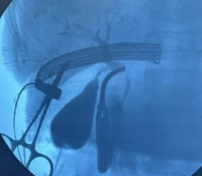
Intraoperative Cholangiography (IOC): This remains the gold standard for conclusive diagnosis [Picture-6]. It is performed under general anaesthesia, with contrast injected into a remnant bile duct (or gallbladder remnant if present) to determine the patency of the extrahepatic biliary tree. Laparoscopic surgical cholangiography is a minimally invasive procedure for identifying biliary atresia.
Diagnostic workup
- Tanget al.ActaPaediatrTaiwan,2007
| *Sn= sensitivity | •Mittalet al.AJR Am JRoentgenol,2011 |
|---|---|
| •Leeet al.JPediatrSurg.2016 | |
| Sp= specificity | •Mizutaet al.JPediatr.2015 |
Picture-4: Depiction of Diagnostic Work-up.
Percutaneous Liver Biopsy: A percutaneous liver biopsy, which exhibits distinctive characteristics of bile duct proliferation, portal tract enlargement and fibrosis, and bile clogging, is extremely accurate (around 95%) in distinguishing BA from other causes such as newborn hepatitis.
Risk prediction scoring system by Redkar R, et al
| Score | Cumulative | Risk | ||
|---|---|---|---|---|
| Score | ||||
| Grade | Grade-1 | 1 | 3 - 7 | Mild |
| of | Grade-2 | 2 | ||
| Fibrosis | Grade-3 | 3 | 8 - 16 | Moderate |
| Grade-4 | 4 | |||
| Grade-5 | 10 | 17 - 20 | Severe | |
| Grade-6 | 12 | |||
| Ratio of | 0 – 1 | 1 | Based on the scores, the authors | |
| AST/ALT | 1.01 -2.19 | 2 | recommend: | |
| 2.20 – 2.39 | 3 | 1. Between 3 and 7 | : Should | |
| >2.4 | 4 | perform a Kasai | ||
| APRI Index | 0- 1 | 1 | Portoenterostomy (KPE) | |
| 1.01-1.8 | 2 | |||
| 1.81-2.39 | 3 | |||
| 2. Between 8 and 16: | Could | |||
| >2.4 | 4 | |||
| recommend a KPE if the child is | ||||
| not fit for a liver transplant |
3. Between 17 and 20: Should recommend a liver transplantation
AST- Aspartate transaminase, ALT- Alanine transaminase, APRI- Aspartate platelet ratio index
Management of Biliary Atresia
The Kasai hepatic portoenterostomy (HPE) surgery is the main surgical intervention. In order to allow bile to drain straight into the gut, the surgeon eliminates the atretic extrahepatic bile duct remains and joins a loop of the baby's intestine to the raw liver surface at the porta hepatis. Bile outflow from the liver's surface into the gut is made possible by this operation.
Kasai Hepatic Portoenterostomy Procedure (KPE)
KPE is carried out by attaching the proximal portal plate to a Roux-en-Y jejunal loop and removing the obliterated extrahepatic fibrous biliary tissue at the porta-hepatis. Following a little incision for intraoperative cholangiography, the results are used to determine the course of surgery. The dissection of the fibrous biliary tissue should reach the point where the umbilical vein joins the left portal vein on the left side and the entrance point of the anterior branch of the right hepatic artery on the right side if the diagnosis of BA is confirmed. A broad raw surface for anastomosis is ensured by carefully cutting the fibrous cone up to the posterior surface of the portal vein. Avoid penetrating the liver parenchyma by limiting the depth of dissection to the liver capsule (above Glisson's capsule). The bile ductules must not be harmed by excessive cautery usage. To avoid cholangitis, a Roux-en-Y limb for porto-jejunal anastomosis should be made at least 40 cm. To determine the extent of fibrosis, a liver biopsy is necessary. Before closure, an abdominal drain is positioned in the subhepatic area. A successful diagnosis and portoenterostomy can also be accomplished using minimally invasive laparoscopic/robotic assisted methods.
Postoperative Care
- Nasogastric Drainage for 48 hours.
- Intravenous Antibiotics for 7-14 days followed by cyclical oral antibiotics for 6 weeks to 3 months.
- Abdominal drains are typically removed before discharge.
- Antibiotics and Choleretics: Prolonged use to prevent postoperative cholangitis.
- Ursodeoxycholic Acid (UDCA): Administered at a dose of 10–15 mg/kg/day.
- Fat-Soluble Vitamins: Vitamin ADEK drops (1 ml/day) and oral or injectable Vitamin K for long-term use.
- *Prednisolone: Given at a dose of 2 mg/kg/day, divided into two doses, with gradual tapering over 2–3 weeks (Author do not use steroids routinely).
Adjunctive medical therapy includes things like steroids and ursodiol. Support from nutrition is crucial. Medium-chain triglycerides (MCT) are frequently employed in high-calorie formulae since they don't need bile salts to be absorbed. Fat-soluble vitamins (A, D, E, and K) must be taken as supplements. Ursodeoxycholic acid (UDCA) is frequently used to lessen bile acid toxicity and increase bile flow. In cases of extensive cirrhosis or when Kasai fails, a liver transplant is the last resort.
Post-operative Complications
| Early Complications | Prolonged Ileus | Bleeding Cholangitis Bile leak Biliary lake formation and persistent | Late Complications Recurrent Cholangitis Cessation of bile flow | Ascites cholangitis |
|---|---|---|---|---|
| Adhesive bowel obstruction | Persistent Ascites | Hepatopulmonary Syndrome | Portal Hypertension | |
| Secondary bacterial peritonitis | Cirrhosis |
Survival, Prognosis and Long-Term Outcome Following Kasai Portoenterostomy
Late presentation of biliary atresia is quite common in Indian continent. Nearly two thirds of individuals may experience portal hypertension and progressive liver fibrosis even with good postoperative bile flow. Ascites, hypersplenism, and esophageal varices are possible consequences. The most significant prognostic factor described in the literature is the timing of the Kasai PE procedure. Success rates (measured by the clearance of jaundice) are considerably higher when the procedure is performed before 45 to 60 days of life, but survival rates and successful KPE also depend on several other factors too.
Survival rate and successful KPE depends on- Initial Good Biliary Drainage and Sustained Bile Flow: Approximately half of patients who achieve good bile drainage (ranging from 25% to 86%) maintain sustained bile flow (30%- 35%) and are free of long-term complications, whereas one-third (35%) of patients (half of those achieving good bile drainage) will experience the gradual onset of progressive liver failure, with jaundice in the long run.
Prognosis depends on the serum bilirubin level at 3 months after surgical intervention. Types I and II Biliary Atresia generally have a good prognosis when treated early before 60 days while Type III Biliary Atresia having larger bile ductules at the porta hepatis (greater than 150 mm in diameter) is associated with a better prognosis. Syndromic Biliary Atresia and Cytomegalovirus (CMV) Infection associated biliary atresia: tend to have worse outcomes, including difficulty in clearing jaundice and higher overall mortality rates. The prognosis for biliary atresia is still uncertain. Many patients experience problems like portal hypertension and ascending cholangitis, a bacterial infection of the liver, even after a successful Kasai surgery. By adolescence or early adulthood, the majority (around 80%) will need a liver transplant. The only effective treatment for end-stage liver disease brought on by untreated BA and numerous other serious causes of newborn cholestasis is liver transplantation.
Liver Transplantation in Biliary Atresia
About 70–80% of patients eventually need a liver transplant due to biliary atresia, which is the most common cause of liver transplantation in children. After a portoenterostomy, 15– 30% of patients will never experience bile drainage and would eventually develop liver failure, necessitating an early liver transplant. In patients with postoperative biliary atresia, the primary rationale for liver transplantation is the total lack of bile drainage, which results in unavoidable clinical decline. Patients who have worsening liver failure, unsuccessful portoenterostomy, or are referred for surgery later in the course may be candidates for liver transplantation.
Cholangitis, hepatopulmonary syndrome, and pulmonary hypertension—which can result from intrapulmonary shunting—are other possible side effects.
Suggested Readings
1. Coran AG, Caldamone A, Scott Adzich N, Krummel TM, Laberge JM, editors. Pediatric surgery. 7th ed. Oxford: Elsevier Limited; 2012.
2. Moyer, V. A., Balistreri, W. F., Britton, J. C., Schiff, L. Guideline for the evaluation of cholestatic jaundice in infants: recommendations of the North American Society for Pediatric Gastroenterology, Hepatology and Nutrition. Journal of Pediatric Gastroenterology and Nutrition, 2004; 39(2), 115–128.
3. Feldman, A. G., Sokol, R. J. Neonatal Cholestasis: Emerging Molecular Mechanisms, Etiology, and Therapeutics. Journal of Pediatric Gastroenterology and Nutrition, 2013; 57(2), 151–159.
4. Consensus report on neonatal cholestasis syndrome. Pediatric Gastroenterology Subspecialty Chapter of Indian Academy of Pediatrics. Indian Pediatr. 2000; 37:845- 51.
5. Arab, J. P., Karpen, S. J., Dawson, P. A. Bile acid physiology and alterations in cholestasis. Hepatology, 2017; 65(1), 350–362.
6. Tang KS, Huang LT, Huang YH, Lai CY, Wu CH, Wang SM, et al. Gamma-glutamyl transferase in the diagnosis of biliary atresia. Acta Paediatr Taiwan. 2007; 48(4):196- 200.
7. Mittal V, Saxena AK, Sodhi KS, Thapa BR, Rao KL, Das A, Khandelwal N. Role of abdominal sonography in the preoperative diagnosis of extrahepatic biliary atresia in infants younger than 90 days. AJR Am J Roentgenol. 2011;196(4): W438-45.
8. Gu YH, Yokoyama K, Mizuta K, Tsuchioka T, Kudo T, et al. Stool color card screening for early detection of biliary atresia and long-term native liver survival: a 19-year cohort study in Japan. J Pediatr. 2015;166(4):897-902.e1.
9. Li, M. H., Lee, H. C., Tan, H. L., Wu, T. C. (2007). Neonatal jaundice and biliary atresia: a screening program in Taiwan. Journal of the Formosan Medical Association, 106 (Suppl 2), S147–S153.
10. Park, W. H., Choi, S. O., Lee, H. J., Kim, S. T. The triangular cord sign in biliary atresia: an ultrasonographic finding. Journal of Pediatric Surgery, 2000; 35(10), 1406–1408.
11. Tiao, G. M., de Lijster, M. S., Hawkins, H., Finegold, M. Liver biopsy analysis in biliary atresia. Seminars in Pediatric Surgery, 2012, 21(3):171–181.
12. Redkar R, Raj V, Chigicherla S, Tewari S, Tampi C, Joshi S. Risk prediction scoring system to predict the postsurgical outcomes of biliary atresia. J Indian Assoc Pediatr Surg. 2020;25(5):280–5.
13. Kumar T; Kumar B; Yadav R; Poddar U; Upadhayaya VD; Mandelia A, et al.
Percutaneous Transhepatic Cholecysto-Cholangiography: Feasibility, Safety, and Diagnostic Accuracy in Cholestatic Jaundice – A Prospective Study. Journal of Indian Association of Pediatric Surgeons 30(6): p 728-734, Nov–Dec 2025.
14. Redkar R, Agarwal P, Raveenthiran V, Gosavi SJ, Bangar A. IAPS textbook of pediatric surgery. Jaypee Brothers Medical Publishers; 2020a