
Introduction
Gastroschisis is one of the spectrum of congenital abdominal wall defects.1 Gastroschisis is usually less than 4cm in diameter, with no covering membrane and the contents float within the amniotic fluid. The contents are usually the midgut with stomach and sometimes a gonad. It is almost always to the right side of the umbilical cord and does not involve the umbilicus.1, 2 The other anomalies of the spectrum of congenital abdominal wall defects include umbilical defects with intact rectus abdominis muscles like Omphalocele, pentalogy of Cantrell, ectopia cordis thoracis, cloacal exstrophy, hernia of umbilical cord and umbilical hernia.1 The absence of a sac and the typical right-sided location in gastroschisis differentiates it from the other abdominal wall defects.
At birth, the protruding bowel may appear normal, but with passage of time, usually after 20 minutes of birth, the extruded intestine will get thickened and covered with fibrinous exudate and get matted together and the individual loops cannot be then distinguished from each other.1 Gastroschisis has been reported to be associated with gastrointestinal conditions like atresia, perforation, necrosis or volvulus. Such association has been termed as complicated gastroschisis and has been associated with poor prognosis.1
Epidemiology
1) Global Incidence and Temporal Trends
The global incidence of gastroschisis ranges from 2 to 5 per 10,000 live births, with marked geographic variation.3, 4, 5 Since the late 20th century, many high-income nations of North America, Europe, and Australasia have reported a significant increase in incidence, from the 1980s through the early 2010s, particularly among infants born to young mothers (<20 years). 3, 4, 5 More recent registry data from high-income countries suggest plateauing or modest declines in incidence since approximately 2016, potentially reflecting changes in maternal risk-factor exposure, including reduced smoking prevalence.6, 7 Despite these temporal shifts, young maternal age remains the strongest and most consistent epidemiologic risk factor worldwide.3, 4, 5
2) Epidemiology in India
In contrast to countries with established congenital anomaly surveillance systems, reliable population-based epidemiologic data on gastroschisis in India are lacking. Published Indian literature consists predominantly of hospital-based case series from tertiary neonatal surgical centres, which provide valuable clinical insights but are inherently limited by referral bias and incomplete case ascertainment.8, 9 Recent published reports suggest incidence of approximately 2–5 per 10,000 live births.10 The recent increase might be attributed to improvements in antenatal diagnosis, referral networks, increase in premature deliveries and better survival of preterm babies.10, 11, 12
Associated Risk Factors
Epidemiologic studies consistently identify several risk factors as follow1, 10:
i) Demographic Risk Factors:
- Young maternal age
- Low socioeconomic status
- Poor maternal prenatal care
- Primigravida status
ii) Nutritional factors:
- Low levels of glutathione and a-carotene
- High levels of nitrosamines
- Absence of supplemental vitamin intake during pregnancy
- Absence of folic acid fortification
- Poor nutrition (Maternal obesity has actually been found to be protective)
iii) Most studies show no familial or genetic risk factors
iv) Environmental and other risk factors:
- Living near chemical plants, farms, landfills
- Use of vasoconstrictive agents and drugs like cocaine, methamphetamine, and marijuana during pregnancy
- Cigarette smoking
- Alcohol use
Maternal Age Distribution: Amongst the above stated risk factors, a consistent and striking epidemiologic feature seen in gastroschisis is the strong association with young maternal age. Mothers younger than 20 years have a 5–10-fold higher risk compared with women aged 25– 29 years. The risk decreases progressively with increasing maternal age, suggesting an environmental or behavioural contributors.3, 4, 12
Sex Distribution: Most studies report a slight male predominance, although the difference is small and not always statistically significant.4, 10, 13These associations support a multifactorial etiology, likely involving vascular disruption during early embryogenesis.1, 3, 10, 14, 15 Associated Anomalies and their impact on outcomes: Unlike omphalocele, gastroschisis is usually an isolated anomaly, with chromosomal abnormalities being rare (<2%). However, intestinal complications such as atresia can occur in 30% patients.10 Other systems are rarely involved. Advances in neonatal intensive care and surgical management have improved survival to >90% in high-income countries, though outcomes remain poorer in low-resource settings.1, 10, 13, 15
Clinical Features of Gastroschisis
The defect in gastroschisis is small (usually less then 4cm) and is almost always to the right side of umbilicus (Figure 1). Sometimes, a skin bridge is present between the defect and the umbilicus. The small bowel protrudes through the defect with no covering and is directly exposed to the environment. Sometimes, stomach, large bowel and gonads may be seen protruding. On the other hand, omphalocele is the defect of the umbilicus and the herniating bowel is covered with sac (Figure 2).
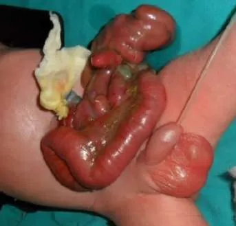
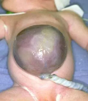
At birth, the protruding bowel may appear normal, but with passage of time, usually after 20 minutes of birth, the extruded intestine will get thickened and covered with fibrinous exudate and get matted together and the individual loops cannot be then distinguished from each other.1 Gastroschisis has been reported to be associated with gastrointestinal conditions like atresia, perforation, necrosis or volvulus. Such association has been termed as complicated gastroschisis and has been associated with poor prognosis.1
The difference between Gastroschisis and omphalocele is tabulated in Table 1.
| S.No. | Features | Gastroschisis | Omphalocele |
|---|---|---|---|
| 1 | Embryologic defect | Failure of closure of the abdominal wall (para-umbilical defect) | Failure of midgut return into abdominal cavity |
| 2 | Location of defect | Mostly to the right of umbilicus | Midline at the umbilical ring |
| 3 | Covering sac | Absent – bowel is directly exposed to the environment | Present (composed of amnion + Wharton’s jelly + peritoneum) |
| 4 | Contents | Usually small intestine ± stomach/colon; sometimes gonads | Bowel ± liver and other viscera |
| 5 | Umbilical cord insertion | Normal, separate from defect | Inserts onto the sac |
| 6 | Bowel appearance | Thickened, edematous, inflamed with “peel” | Usually normal in appearance |
| 7 | Associated anomalies | Rare (intestinal atresia might be present) | Common (cardiac, neural tube, chromosomal) |
| 8 | Chromosomal association | Uncommon | Frequent (Trisomy 13, 18, 21; Beckwith–Wiedemann) |
| 9 | Gestational age / birth weight | Often preterm, low birth weight | Usually term or near-term |
| 10 | Antenatal maternal AFP | Markedly elevated | Mild to moderate elevation |
| 11 | Risk of infection and sepsis | High due to exposed bowel | Lower (intact sac unless ruptured) |
| 12 | Immediate neonatal issue | Fluid, heat loss; bowel injury | Risk of sac rupture |
| 13 | Management at birth | Bowel protection + primary or staged closure (silo) | Sac protection; staged closure if giant omphalocele |
| 14 | Prognosis | Generally good if isolated | Depends on associated anomalies |
Embryogenesis of Gastroschisis
Multiple theories for embryogenesis of gastroschisis have been proposed, but none of them are well-accepted. To understand the embryogenesis of gastroschisis, it is essential to understand the embryology during the organogenesis period. At 3 weeks’ gestation, the four folds develop into the embryonic disk which enclose the body cavities - two lateral folds, one cephalic fold and the caudal fold. Simultaneously, the gut tube is formed along the length of embryo and its elongation and development take place within the umbilical coelom till about10 weeks, after which the gut returns from the space within the umbilical stalk to the peritoneal cavity and undergoes rotation and fixation.1
With the above background comes the first theory of its embryogenesis, i.e., failure of development of umbilical coelom. The elongating intestine then has no space in which to expand and the gut then ruptures out of the body wall to the right of umbilicus, which is relatively unsupported because of resorption of the right umbilical vein at about 4 weeks’ gestation.
Inadequate mesoderm formation has also been proposed which leads to absence or weakness of the muscular and connective tissue layers, producing a paraumbilical wall defect through which abdominal contents herniate.16 An alternate explanation is that the yolk sac and associated vitelline structures fail to incorporate into the umbilical cord, hereby allowing the midgut to exit the abdomen at the point they exit. This theory also seems reasonable as gastroschisis has no covering membrane.1 Another theory of embryogenesis of gastroschisis is abnormal or premature regression of the right umbilical vein, leading to localised ischemia and weakening of the right periumbilical wall and causing gastroschisis.17, 18
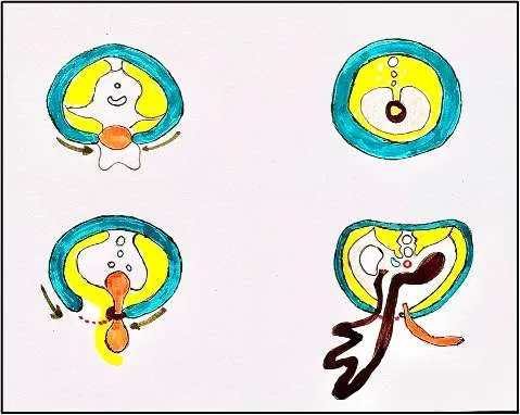
It has also been proposed that gastroschisis results from defective lateral body folding, specifically failure of the right lateral fold to advance and fuse properly.16, 19 The resulting incomplete closure of the abdominal wall leaves a persistent paraumbilical opening (Figure 3). This theory aligns with the anatomic timing of abdominal wall formation during weeks 4–5 of gestation.
The most widely accepted and leading hypothesis is that gastroschisis results from a vascular accident, typically involving the right omphalomesenteric artery or right umbilical vein, leading to ischemic injury and subsequent full-thickness disruption of the abdominal wall.16, 18 This also explains the defect's consistent right-sided location and is supported by the frequent association of bowel injury and atresia, which are compatible with an ischemic mechanism. Recent studies suggest that the above stated embryologic pathways might be modified or triggered by external teratogenic factors—especially vasoactive exposures such as smoking, young maternal age, NSAIDs, and certain recreational drugs.20, 21 These factors might increase the susceptibility to vascular disruption or interfere with mesodermal or umbilical ring development. In a comprehensive review of the literature gastroschisis was reported to be induced by 22 teratogens.1
The appearance of the bowel is most often a postnatal event. At the moment of birth, the bowel in gastroschisis is usually quite normal. Twenty minutes later it begins to acquire the characteristic changes. These changes may be due to exposure to air, but more likely they are related to mesenteric venous occlusion at the level of the abdominal wall defect with resultant edema and transudation of proteinaceous fluid.1
Recent systematic evidence (2023–2025) on etiopathogenesis of gastroschisis
Several recent systematic reviews reported a multifactorial model vascular susceptibility along with environmental triggers, possibly modified by genetic susceptibility in the etiopathogenesis of gastroschisis, as follows14, 22, 23, 24, 25:
- Genetics/genomics: A recent systematic review by Marquart JP et al concluded that though the current human genetic studies are limited, they point toward gene–environment interactions for gastroschisis etiology; more modern omics/epigenetic studies are recommended.22
- Experimental (animal) models: Another systematic review of fetal lamb experiments by Arai T et al summarized the different surgical/experimental methods which induce simple versus complex gastroschisis. They tried to clarify prenatal manipulations which produce ischemic/structural bowel changes.23
- Infectious exposures (GUTI): The systematic review and meta-analysis by Vinogradov R et al concluded that periconceptional genitourinary infections (UTI and STI) might be associated with increased gastroschisis risk, supporting environmental/vascular-mediated mechanisms.24
- Medication exposures/teratogens: Baldacci S et al, published a recent meta-analysis on medication use in pregnancy and reported associations between first-trimester exposure to several over the counter drugs like aspirin, ibuprofen, pseudoephedrine/phenylpropanolamine and increased risk of gastroschisis, again consistent with environmental/vascular or teratogenic contributors to embryogenesis.14
Antenatal Diagnosis, Monitoring, and Management
Accurate prenatal diagnosis is important so that planning of the management and counselling of to be parents could be done well.
Ultrasonography: Antenatal ultrasonography, especially the second-trimester anomaly scan (18–22 weeks), is the commonest diagnostic modality used. Although first-trimester diagnosis is now been increasingly reported with advances in imaging technology. The characteristic sonographic features include free-floating bowel loops herniating through a right-sided paraumbilical abdominal wall defect, absence of a covering membrane, and a normal insertion of umbilical cord.13, 16, 20, 26
Other markers: Alpha-fetoprotein in both maternal serum and amniotic fluid and Acetylcholine esterase in amniotic fluid are elevated in abdominal wall defects; provided open neural tube defects have been ruled out.
Gastroschisis can be differentiated from omphalocele at ultrasound by its paraumbilical location and lack of a covering membrane. Presence of liver and large size of the defect would help to differentiate ruptured omphalocele from gastroschisis. Sensitivity of ultrasound and maternal serum Alpha-fetoprotein screening is close to 80% in diagnosing abdominal wall defects10
Antenatal Counselling and Monitoring: Counselling of the to be parents about the nature of primary defect, associated anomalies and genetic syndromes. After the associated lethal defects have been ruled out, the pregnancy is to be carried till term with close monitoring and surveillance as follows:
a) Serial Ultrasound Surveillance: After diagnosis, serial ultrasound surveillance is recommended every 2–4 weeks, increasing to weekly or biweekly in the third trimester. Monitoring of the fetus is to be done for fetal growth, amniotic fluid volume, and bowel characteristics, including dilatation, wall thickness, and echogenicity.
b) Fetuses with gastroschisis are at increased risk of fetal growth restriction and stillbirth, antenatal surveillance should include umbilical artery Doppler studies, as well as non-stress testing or biophysical profiles from 32–34 weeks’ gestation 10, 27, 28, 29 c) Parents should be encouraged to follow-up and get delivery done at a tertiary care centre with well-equipped NICU, paediatric surgery and neonatal anaesthesia availability
A recent systematic review and meta-analysis (2023) demonstrated that intra-abdominal bowel dilatation is significantly associated with complex gastroschisis, including bowel atresia, necrosis, and perforation, whereas isolated extra-abdominal bowel dilatation has limited predictive value.27 Timing and mode of Delivery: Current evidence supports expectant antenatal management with close surveillance rather than routine early preterm delivery. A systematic review and meta-analysis by Morris et al30 showed that elective delivery before 36 weeks does not reduce mortality and increases neonatal respiratory morbidity.30
The optimal timing of delivery remains controversial. Recent systematic reviews and individual participant data meta-analyses suggest that delivery at 37–38 weeks’ gestation provides the best balance between the risks of stillbirth and prematurity in uncomplicated gastroschisis.30, 31 Planned early preterm delivery has not demonstrated a survival benefit.¹¹
Multiple systematic reviews and meta-analyses consistently demonstrate no advantage of elective caesarean section over vaginal delivery in terms of neonatal survival, bowel outcomes, or length of hospital stay32, 33 Accordingly, vaginal delivery is recommended32; however, the mode of delivery should be best decided by the treating Obstetrician and should be as per the obstetric indications
Antenatal Interventions: Antenatal corticosteroids can be administered according to routine obstetric indications for threatened preterm birth; there is no evidence of a gastroschisis-specific benefit.30 Experimental approaches such as amnio-exchange or in-utero bowel covering remain investigational and have not recommended outside clinical trials.34
Summary of evidences as per the recent Randomized Controlled Trials
- RCTs done were small and underpowered, limiting definitive conclusions.35-38
- Elective early preterm delivery (34–36 weeks) has been tested but does not improve outcomes and may increase complications (e.g., sepsis). Early elective delivery hence is not recommended solely to improve gastroschisis outcomes.
- There is no established prenatal intervention from randomized trials that alters the course of gastroschisis. Amnio-exchange has not been shown to have benefit in reducing bowel-related morbidity.
- A large ongoing RCT (GOOD Study) has been designed to address the gaps in evidence on delivery timing, but the results are still pending38
Postnatal Management
As stated earlier, delivery of such babies is recommended at a tertiary care centre with well-equipped NICU, paediatric surgery and neonatal anaesthesia availability. Rapid transfer of the neonate from delivery room to the neonatal operating room increases the chances of primary closure of the defect.1
The main principles of management are:
- Stabilization of neonate
- Reduction of viscera
- Closure of the defect
- Management of the associated anomalies (if any)
Immediate Postnatal Management
Stabilization of the neonate: It begins in the delivery room itself.
Protection of the exposed bowel: The exposed bowel should be immediately protected using a sterile warm saline-soaked gauze or sterile urobag, to minimize evaporative heat loss and bowel dilatation. It should be ensured that the defect in gastroschisis is not very narrow to compromise the bowel vascularity of the exposed bowel. The neonate should be positioned with support to the bowel to prevent traction on the mesentery.10, 13 Orogastric tube placement, catheterization and per rectal wash are recommended to decompress the gut and reduce bowel distension and aspiration risk.10
Thermoregulation and Fluid Management: Infants with gastroschisis are at high risk of hypothermia and third-space fluid losses. Aggressive fluid resuscitation (150 – 175ml/kg/day ) is required during the first 24 – 48 hours, guided by urine output, serum electrolytes, and acid–base status.1, 10, 13, 39
Respiratory Support and Antibiotics: Respiratory support should be provided as needed. Broad-spectrum intravenous antibiotics should be initiated early to reduce the risk of sepsis related to bowel exposure, typically continued until definitive abdominal closure and clinical stabilization.
Surgical Management
1) Reduction and Primary Closure
- Without mesh: The surgical goal is safe reduction of herniated viscera and abdominal wall closure without causing abdominal compartment syndrome. This can be accomplished either without prosthetic mesh in selected neonates with minimal bowel edema and adequate abdominal domain.
- With mesh: Closure with mesh may result in complications and require removal later
Steps of primary closure
- The bowel contents are reduced gradually; this can be aided by decompressing the bowel by gastric aspiration, rectal washes and milking.
- The abdominal wall may be gently stretched by finger posteriorly to anteriorly to prevent closure under tension
- If still the closure feels tight, then relaxing incisions can be given in the abdominal wall fascia
- The closure should be done using interrupted sutures.
- It is important to ensure that the primary closure is not under tension which can cause abdominal compartment syndrome. The following parameters, if present, should prompt the surgeon to apply silo instead of primary closure10:
- Mean airway pressure more than 25cm of water
- Intravesical pressure more than 20cm of water
- Saphenous intravenous line unable to flow by gravity
Even after primary closure, the neonate should be monitored post-operatively for early signs of abdominal compartment syndrome like persistent tachycardia, oliguria, increased ventilator setting requirements and tensed abdomen. Such patients might need decompression with silo placement.
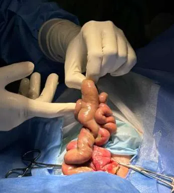
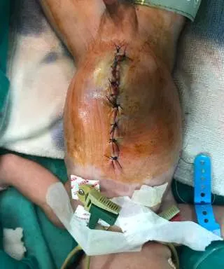
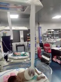
2) Gradual/Staged Reduction: This is done using a silo and is preferred when bowel edema is significant or primary closure would compromise ventilation or perfusion.40, 41, 42 This can be applied bed side and kept tied to keep the bowel in place and tightened gradually to cause gradual reduction without compromising the ventilation. When the complete eviscerated bowel can be reduced, then primary closure can be done.
3) Creation of ventral hernia with delayed repair: In this technique, only the skin is closed after reduction of the bowel at neonatal period and the resulting ventral hernia is repaired later when the child grows. This might be difficult and may require prosthesis or tissue expanders as the bowel grows into the defect and ventral hernia becomes larger and difficult to reduce.
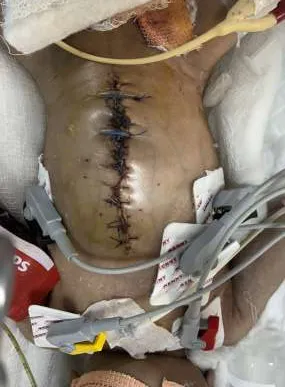
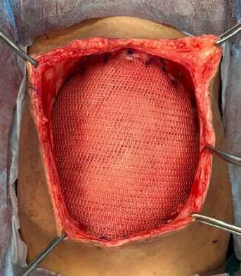
Multiple systematic reviews have demonstrated no difference in survival between primary closure and silo placement, although silo use may reduce ventilatory compromise and improve physiologic stability.11, 41 Management of Complex Gastroschisis: Approximately 10–20% of infants have complex gastroschisis, characterized by intestinal atresia, necrosis, perforation, or volvulus.43 These infants often require:
- Delayed definitive surgery
- Resection of nonviable bowel
- Prolonged parenteral nutrition
- Increased risk of short bowel syndrome and mortality43, 44
Postoperative and Nutritional Management
1. Parenteral and Enteral Nutrition: Total parenteral nutrition (TPN) is initiated early and continued until bowel function recovers. Enteral feeds are introduced gradually, usually after resolution of ileus and reduction of gastric aspirates 40 Early trophic feeding, when feasible, should be encouraged as it is associated with shorter hospital stay and earlier achievement of full enteral nutrition.45
2. Infection and Complication Surveillance 3. Management of post-operative bowel dysmotility: This poses a difficult challenge as bowel motility may take long to recover. Early gradual trophic feeds should be encouraged as tolerated. Use of prokinetic drugs like domperidone, erythromycin and metoclopramide have been tried with variable success.
4. Infants require close monitoring for:
- Central line–associated bloodstream infections
- Necrotizing enterocolitis-like bowel injury
- TPN-associated cholestasis
- Abdominal compartment syndrome40
Management of Cryptorchidism
This condition is associated with gastroschisis with an incidence of 15-30%. The herniated testis should be replaced in the abdominal cavity at the time of first surgery as this results in normal descent of the testis into the scrotum in majority of the cases.
Hospital Outcomes
Survival rates for simple gastroschisis exceed 90–95% in high-resource settings. However, morbidity remains substantial, particularly in complex cases, with prolonged hospitalization and feeding difficulties being common.11, 43, 44 Major causes of death are prematurity, sepsis and ischemia of the bowel.
Long-Term Follow-up
1. Growth and Nutrition: Long-term follow-up focuses on growth, nutritional adequacy, and gastrointestinal function. While most survivors achieve normal growth by early childhood, infants with complex gastroschisis are at increased risk of:
- Growth failure
- Feeding intolerance
- Intestinal dysmotility44, 45
2. Neurodevelopmental Outcomes: Most children with uncomplicated gastroschisis have normal neurodevelopmental outcomes, although prematurity and prolonged hospitalization may contribute to subtle delays.46, 47 Routine developmental surveillance is recommended during infancy and early childhood.
3. Gastrointestinal and Surgical Sequelae: Late complications may include:
- Adhesive small bowel obstruction
- Incisional hernia
- Chronic abdominal pain or constipation48, 49
Lifelong follow-up is usually not required for simple gastroschisis unless for children with complex disease or short bowel syndrome.
Conclusion/Summary
Prognosis of antenatally detected gastroschisis remains good in institutes with well-equipped and co-ordinated multidisciplinary neonatal, surgical, and nutritional care. Early stabilization, appropriate surgical strategy, meticulous postoperative management, and long-term follow-up are essential to optimize survival and functional outcomes.
Emerging Concepts and Research Gaps
Despite extensive observational data, randomized evidence for antenatal interventions remains limited. Systematic reviews conclude that procedures such as amnio-exchange and fetal bowel covering have no proven benefit and should remain experimental.30, 34 Ongoing multicentre randomized trials evaluating optimal timing of delivery are anticipated to address key evidence gaps.
Future Prospects in Gastroschisis
1. Improved Antenatal Risk Stratification: One of the major future goals in gastroschisis care is the development of reliable antenatal predictors of complex disease and adverse outcomes. Future research needs to focus upon:
- Composite prediction models integrating ultrasound parameters (intra-abdominal bowel dilatation, growth restriction), Doppler findings, and gestational age3
- Machine-learning–based risk models using longitudinal imaging and clinical data to better predict complex gastroschisis and neonatal morbidity50
Such tools may be beneficial for individualized counselling, optimized timing of delivery, and targeted neonatal preparedness.
2. Biomarkers and Amniotic Fluid Research: Experimental and translational research suggests that inflammatory mediators in amniotic fluid contribute to bowel damage in gastroschisis.27 Although investigational, the future prospects might focus on:
- Identification of biochemical biomarkers (cytokines, bile acids, oxidative stress markers) to predict bowel injury severity28, 30
- Development of non-invasive maternal serum or amniotic fluid markers to supplement ultrasound findings28, 30
3. Refinement of Antenatal Management Strategies: There are no high-quality randomized evidence guiding antenatal management available yet. Future advances are expected from:
- Large multi-center randomized controlled trials evaluating optimal timing of delivery (e.g., early-term vs expectant management)51
- Standardization of antenatal surveillance protocols based on risk stratification rather than uniform intensive monitoring
4. Innovations in Fetal Therapy: At present, fetal surgical intervention for gastroschisis remains experimental and is limited to research settings.32, 33 Although previous trials of amnio-exchange and fetal bowel covering have not shown benefit,32 future fetal therapies might focus on:
- Targeted modulation of the intra-amniotic inflammatory environment
- Advanced biomaterials or minimally invasive fetal interventions
5. Advances in Neonatal and Surgical Care: Future neonatal management aims to reduce morbidity rather than mortality, which is already low in simple gastroschisis. Anticipated advances include:
- Standardized feeding protocols and enhanced recovery pathways to reduce time to full enteral feeds11
- Improved strategies to prevent intestinal failure–associated liver disease
- Refinement of minimally invasive and bedside closure techniques to reduce ventilator dependence and hospital stay40
6. Long-Term Outcomes and Quality of Life: Future research should also focus on long-term functional outcomes, including:
- Growth, gastrointestinal function, and nutrition
- Neurodevelopmental and psychosocial outcomes
- Health-related quality of life in adolescence and adulthood44, 48
This might be done by establishment of long-term follow-up registries to understand lifelong outcomes.
7. Global Surveillance and Prevention: Future prospects also include strengthening population-based congenital anomaly surveillance, particularly in low- and middle-income countries.¹⁶ Improved data quality may:
- Clarify true global incidence trends
- Enable identification of environmental and behavioural risk factors
- Inform preventive public health strategies
References
- Klein MD. Congenital Defects of the abdominal wall. in: O'Neil JA Jr, Rowe MI, Grosfeld JL, Fonkalsrud EW, Coran AG, editors. Pediatric Surgery. 8th ed. Mosby, New York, USA.2021. p. 973-984.
- Logghe HL, Mason GC, Thornton JG, Stringer MD. A randomized controlled trial of elective preterm delivery of fetuses with gastroschisis. Journal of pediatric surgery. 2005 Nov 1;40(11):1726-31.
- Loane M, Dolk H, Bradbury I; EUROCAT Working Group. Increasing prevalence of gastroschisis in Europe 1980–2002: a phenomenon restricted to younger mothers? Paediatr Perinat Epidemiol. 2007;21(4):363-369.
- Feldkamp ML, Botto LD. Developing a research and public health agenda for gastroschisis: how do we bridge the gap between what is known and what is not?. InAmerican Journal of Medical Genetics Part C: Seminars in Medical Genetics 2008 Aug 15 (Vol. 148, No. 3, pp. 155-161). Hoboken: Wiley Subscription Services, Inc., A Wiley Company.
- Feldkamp ML, Canfield MA, Krikov S, Prieto‐Merino D, Šípek Jr A, LeLong N, Amar E, Rissmann A, Csaky‐Szunyogh M, Tagliabue G, Pierini A. Gastroschisis prevalence patterns in 27 surveillance programs from 24 countries, International Clearinghouse for Birth Defects Surveillance and Research, 1980–2017. Birth defects research. 2024 Feb;116(2):e2306.
- Jones AM. Increasing prevalence of gastroschisis—14 states, 1995–2012. MMWR. Morbidity and mortality weekly report. 2016;65.
- Schuh JM, Cabacungan E, Wagner AJ. Birth Prevalence of Gastroschisis in the United States: 2016-2022. Pediatrics. 2025;155(4):e2024068696. doi:10.1542/peds.2024- 068696
- Gupta R, Gurjar BL. Early outcome analysis of gastroschisis from a high-volume tertiary care institute in India: a prospective observational study. J Neonatal Surg. 20212:20.
- Singh AP. Is the incidence of gastroschisis increasing recently? J Neonatal Surg. 2018;7:25.
- Roy Choudhury S. Pediatric Surgery : A Quick Guide to Decision-Making. Singapore: Springer, 2018. https://doi.org/10.1007/978-981-10-6304-6
- Wright NJ, Sekabira J, Ade-Ajayi N. Care of infants with gastroschisis in low-resource settings. Semin Pediatr Surg . 2018;27(5):321 -326. doi:10.105j.sempedsurg.2018.08.004
- Werler MM, Mitchell AA, Shapiro S. Demographic, reproductive, medical, and environmental factors in relation to gastroschisis. Teratology. 1992;45(4):353-360. doi:10.1002/tera.1420450406
- Bradnock TJ, Marven S, Owen A, Johnson P, Kurinczuk JJ, Spark P, et al. Gastroschisis: one year outcomes from national cohort study. BMJ. 2011;343:d6749.
- doi:10.1136/bmj.d6749
- Baldacci S, Santoro M, Mezzasalma L, Pierini A, Coi A. Medication use during pregnancy and the risk of gastroschisis: a systematic review and meta-analysis of observational studies. Orphanet J Rare Dis. 2024;19(1):31. Published 2024 Jan 30. doi:10.1186/s13023-023-02992-z
- Bradnock TJ, Knight M, Kenny S, Nair M, Walker GM; British Association of Paediatric Surgeons Congenital Anomalies Surveillance System. Gastroschisis: mortality, morbidity, and associations with gestational age at birth. J Pediatr Surg. 2017;52(6):949-955. doi:10.1016/j.jpedsurg.2017.01.005
- Feldkamp ML, Carey JC, Sadler TW. Development of gastroschisis: Review of hypotheses, a novel hypothesis, and implications for research. Am J Med Genet A. 2007;143A(7):639-52. doi:10.1002/ajmg.a.31578
- Stevenson RE, Rogers RC, Chandler JC, Gauderer MWL, Hunter AGW. Escape of the yolk sac: A hypothesis to explain the embryogenesis of gastroschisis. Clin Genet. 2009;75(4):326-3. doi:10.1111/j.1399-0004.2008.01142.x
- Lubinsky M. A vascular and thrombotic model of gastroschisis. Am J Med Genet A. 2014;164A(4):915-7. doi:10.1002/ajmg.a.36370
- Bargy F, Beaudoin S. Comprehensive developmental mechanisms in gastroschisis. Fetal Diagn Ther. 2014;36(3):223-30. doi:10.1159/000360080
- Beaudoin S. Insights into the etiology and embryology of gastroschisis. Semin Pediatr Surg. 2018;27(5):283-8. doi:10.105j.sempedsurg.2018.08.005
- Opitz JM, Feldkamp ML, Botto LD. An evolutionary and developmental biology approach to gastroschisis. Birth Defects Res. 2019;111(6):294 -311. doi:10.1002/bdr2.1481
- Marquart JP, Nie Q, Gonzalez T, Jelin AC, Broeckel U, Wagner AJ, Reddi HV. Genetics and Genomics of Gastroschisis, Elucidating a Potential Genetic Etiology for the Most Common Abdominal Defect: A Systematic Review. J Dev Biol. 2024 Dec 19;12(4):34. doi:10.390/jdb12040034.
- Arai T, Tianthong W, Russo FM, Basurto D, Joyeux L, De Coppi P, Deprest J.
- Experimental induction of complex gastroschisis in the fetal lamb: systematic review. Prenat Diagn. 2024 Oct;44(11):1372–1380. doi:10.1002/pd.6647.
- Vinogradov R, et al. Genitourinary infection and gastroschisis: a systematic review and meta-analysis. Birth Defects Res. 2024 Jul;116(7):e2377. doi:10.1002/bdr2.2377.
- Putri DU, Wibowo YP. Prevalence, management and outcome of gastroschisis: a comprehensive systematic review. J Adv Res Med Health Sci. 2024 Mar;10(3):400–
- doi:10.61841/jghz5x97. (open-access systematic review covering 2014–2024 literature).
- Calvert N, Damiani S, Hiorns MP. Antenatal diagnosis of abdominal wall defects. Prenat Diagn. 2019;39(10):857-866.
- Raitio A, Hijkoop A, et al. Prenatal ultrasound markers and outcome in gastroschisis: a systematic review and meta-analysis. Ultrasound Obstet Gynecol. 20261(2):145-154.
- Tamura M, et al. Prediction of complex gastroschisis: a systematic review. Prenat Diagn. 2020;40(5):53-541.
- South AP, Stutey KM, Meinzen-Derr J. Gastroschisis: epidemiology and antenatal considerations. Clin Perinatol. 2020;47(2):321-32.
- Morris RK, Malin GL, et al. Timing of elective delivery in gastroschisis: a systematic review and meta-analysis. BJOG. 2022;129(1):28-38.
- van der Hoeven MAHBM, et al. Planned early delivery versus expectant management for gastroschisis: an individual participant data meta-analysis. Ultrasound Obstet Gynecol. 20262(3):321-30.
- Al-Kaff A, MacDonald SC, et al. Mode of delivery in gastroschisis: a systematic review and meta-analysis. J Pediatr Surg. 2020;55(4):637-643.
- South AP, et al. Mode of delivery and neonatal outcomes in gastroschisis. Clin Perinatol. 2020;47(2):33-347.
- Deprest J, Gratacos E, et al. Fetoscopic interventions for gastroschisis: current evidence and future directions. Prenat Diagn. 2019;39(9):687-694
- Shamshirsaz AA, Lee TC, Hair AB. Elective delivery at 34 weeks vs routine obstetric care in fetal gastroschisis: randomized controlled trial. Ultrasound Obstet Gynecol. 2019;55(1):15–19.
- A randomised controlled trial of amnioexchange for fetal gastroschisis. BJOG. 2019;126(10):1242–1242.
- Elective preterm delivery of fetuses with gastroschisis: a randomized controlled trial. J Pediatr Surg. 2005;40(11):1726–1731.
- Gastroschisis Outcomes of Delivery (GOOD) Study. ClinicalTrials.gov; NCT02774746. Ongoing RCT comparing 35 weeks delivery vs expectant management.
- Snyder CL. Outcome analysis for gastroschisis. J Pediatr Surg. 2019;54(8):1522-1526.
- Kunz SN, Tieder JS, Whitlock K, et al. Primary fascial closure versus silo placement in gastroschisis. J Pediatr Surg. 20148(4):845-857.
- Zani A, Eaton S, Puri P, et al. International survey on the management of gastroschisis. J Pediatr Surg. 2014;49(3):407-412.
- Skarsgard ED, Claydon J, Bouchard S, et al. Canadian Pediatric Surgery Network outcomes in gastroschisis. J Pediatr Surg. 2015;50(3):431-436.
- Molik KA, Gingalewski CA, West KW, et al. Gastroschisis: a plea for risk categorization. J Pediatr Surg. 2001;36(1):51-55.
- Harris EL, Minutillo C, Hart SJ, et al. The long-term outcomes of gastroschisis: a systematic review. J Pediatr Surg. 2014;49(10):1542-1551.
- Veenma DCM, et al. Early enteral feeding following gastroschisis repair. J Pediatr Surg. 2012;47(10):1896-1901.
- Lap CC, et al. Neurodevelopmental outcomes in children with gastroschisis. Early Hum Dev. 2016;103:81-86.
- Holland AJA, et al. Developmental outcome after gastroschisis repair. J Pediatr Surg. 2010;45(6):1250-1255.
- Fullerton BS, Sparks EA, Morrow KA, et al. Long-term outcomes of children with gastroschisis. J Pediatr Surg. 2017;52(12):1958-1964.
- Emil S, Canvasser N, Chen T, et al. Long-term bowel obstruction following gastroschisis repair. Pediatr Surg Int. 2016;32(9):847-852.
- Bhide P, Gund P, Kar A. Prevalence of congenital anomalies in India: systematic review and meta-analysis. BMC Pediatr. 2016;16:186.
- Sparks TN, Shaffer BL, Feldstein VA, et al. Timing of delivery and outcomes in gastroschisis: meta-analysis. Am J Obstet Gynecol. 2017;216(1):32-39.