
Necrotising Enterocolitis (NEC)is an acquired acute intestinal inflammatory condition of predominantly preterm newborns with significant morbidity and mortality. Although, there is variability in incidence reporting from centers of various geographical regions, 10% is the best approximation of overall incidence and 27%-52% of those need surgical intervention.1 NEC is almost always seen in Very Low Birth Weight (VLBW <1500g) and Extremely Low Birth Weight (ELBW <1000g) newborns with increasing rates of preterm births being cared for in the in NICU. However, approximately 10% occur in full term neonates. It is the most common newborn surgical emergency, and despite advances in the understanding of the condition, it remains a leading cause of morbidity and mortality in the NICU.2
Etiopathogenesis: After decades of investigations, the etiology of NEC appears to be multifactorial. It is a complex combination of intestinal immaturity, enteral feeding in a vulnerable neonate, microbial dysbiosis, immature immune defences and compromised mucosal integrity with local ischemia or hypoxia resulting in an inflammatory cascade leading to necrosis of the intestinal segment.
Three hit model tries to explain the pathogenesis.3
Passive immune deficit: The colostrum and/or breast milk provides the immunoglobulins and innate immune factors to the newborn. The growing foetus also acquires these by active transport across the placenta in the third trimester. Hence the infants born before 30 weeks are in a passive immune deficit state, which can only be partially supplemented by breast milk. This can explain the prevalence and severity of NEC in infants born before or at the threshold of the third trimester. Compromise of the intestinal epithelial barrier appears to be the first event.
Innate immune priming: Repeated episodes of inflammation (due to pathogens or other stimuli) can over-prime the innate immune system which predisposes the neonate to NEC. The cascade controlling the normal surveillance and subsequent pyknosis of invading mucosal pathogens when goes uncontrolled ,leads to abnormal apoptosis and subsequent necrosis. A critical stress or trigger: A third factor that pushes the neonate over the threshold during this period of susceptibility is likely the pathogen. There is almost no known neonatal pathogen (bacterial or viral) that has not been associated with NEC.
NEC may involve a single (in approximately 50%) or multiple discontinuous segments.2 Terminal ileum followed by colon are most commonly involved. The most severe variant (approximately in 19% of operated cases) is the necrosis of more than 75% of the intestine which is termed NEC totalis.
Clinical Features: At the onset, the clinical features are non-specific. the symptoms represent a physiological instability with temperature instability, hypoactivity, recurrent apnea, hypoglycemia, bradycardia and features of shock. The GI symptoms usually include high feed residue, abdominal distension, blood in stools (usually small amounts). As the disease progresses, examination findings of ischemic bowel like abdominal tenderness, palpable bowel mass, and erythematous discoloration of the abdominal wall (necrotic bowel) can be noted. Presentation, diagnosis, and differential diagnoses. In some ELBW neonate, this progress may occur within a matter of 24 hrs. resulting in mortality.
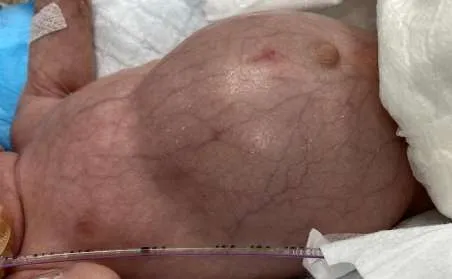
Abdominal distension
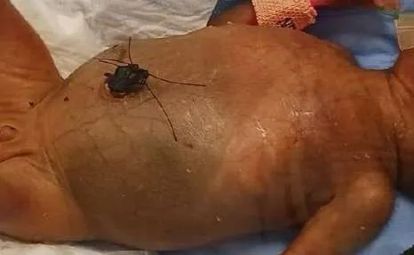
Abdominal wall discoloration Diagnosis: Even after a lot of research, it is still difficult to find a define a diagnosis of NEC. NEC is a spectrum of disease and for which Bell’s staging has been in place to help with prognostication. A very acceptable bedside definition for NEC has been proposed by Dr. Peter Gordon, which he described as Two out of Three rule.4
Patients may be given a diagnosis of Preterm NEC if they have abdominal distension, ileus and/or bloody stools and meet at least 2 of the criteria below:
1.Pneumatosis and/or portal air by ultrasound or abdominal X-ray at presentation 2. Persistent platelet consumption (Platelet count <150,000/µL persisting for ≥3 days)
3. Post-menstrual age (PMA) at disease onset is more consistent with NEC than spontaneous intestinal perforation (SIP)
Bell’s Staging
Bell Stage 1: Suspected disease Mild systemic sigs-bradycardia, apnea, temperature instability Mild intestinal signs-abdominal distention, emesis, bloody stools Non-specific or normal abdominal radiographs Bell Stage 2:Definite disease
Mild to moderate systemic signs Additional intestinal signs-absence of bowel sounds, abdominal tenderness Radiographic signs-pneumatosis intestinalis and/or portal venous gas Laboratory derangements-metabolic acidosis, thrombocytopenia Bell Stage 3: Advanced disease (Surgical NEC)
Severe systemic signs-respiratory failure, hypotension Severe intestinal signs-abdominal discoloration, severe distention Severe radiographic signs-pneumoperitoneum Severe laboratory derangements-disseminated intravascular coagulopathy, severe respiratory and metabolic acidosis
Differential Diagnosis
1.Sepsis in a newborn also presents with similar features at the onset.
2.Spontaneous Intestinal Perforation (SIP): This is a condition with a single bowel wall perforation typically occurring in the terminal ileum in a premature newborn. Although not always possible to differentiate clinically from Surgical NEC, but some of the differentiating features include:
In general, it occurs in more premature babies when compared to NEC Occurs in significantly more premature and lesser weight babies than NEC.
Occurs earlier than NEC; median age for SIP is 7 days, whereas for NEC is 14 days
Investigations
Laboratory: Usually neutropenia (and seldom neutrophilia), thrombocytopenia and metabolic acidosis is found in the infants with NEC. Persistent thrombocytopenia is nearly always present in these newborns.
C-Reactive Protein being elevated more than 10mg/L for hrs.
Occult blood in the stool is positive in the NEC babies although it is not specific for it. Intestinal Fatty acid-binding protein (I-FAB) is a small cytoplasmic protein of the mature enterocytes. Enteric cell death leads to the release of I-FAB in circulation. However, it is convinient to measure its level in urine of the newborn as it passes through the glomerulus. Fecal Calprotectin is also a marker of inflammation of the bowel. Both I-FAB and calprotectin serve as indicators of disease severity rather than as a screening tool for NEC.
Blood Culture is positive in approximately 30–40% of cases. Common organisms: Gram-negative bacilli and anaerobes . Negative culture does not exclude NEC.
Imaging
Plain X ray abdomen: Supine view will show dilated bowel loops and lateral decubitus may show air-fluid levels as non-specific finding in NEC.
Pneumatosis intestinalis (gas within the layers of bowel wall) is also a non- specific finding in NEC reported in 19-98% of the cases1 (it can also be seen in infants with enterocolitis of Hirschsprung disease, inspissated milk syndrome, pyloric stenosis, severe diarrhoea, carbohydrate intolerance). It is usually an early rather than late sign and it may be fleeting occurrence.
Portal venous Gas (PVG) is seen as branching radiolucent shadows over the liver. It is seen in about 30% of the cases, and again it is a fleeting sign. It indicates a severe disease in majority of the cases.
Pneumoperitoneum: Free air in the peritoneal cavity can be seen in supine film as a ‘football’ sign (diffuse round radiolucency like a football in upper abdomen) or as ‘double wall’ (air both within and outside the bowel wall). In a lateral decubitus film, it is distinctly seen as hyperlucent shadow over the liver surface. (Radiograph). It is a sign of perforation of the bowel but seen only in around 60% of the perforations in NEC.
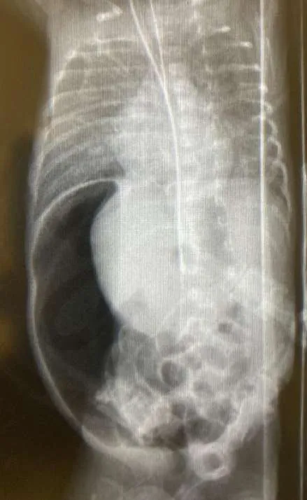
Pneumoperitoneum in a lateral decubitus film
Fixed Bowel loop: In this a single loop or several loops of dilated bowel remain unchanged in position and configuration for 24 to 36 hours in an abdominal plain radiograph. Although considered by some but It is not always indicator of gangrenous bowel. Ultrasound (US): This modality has been found superior to plain X ray in demonstrating PVG, echoic free fluid (EFF) or focal fluid collections . Additionally it can also detect decreased bowel wall perfusion . EFF observed on AUS is an early sign of intestinal perforation and hence, it tip the balance in favour of surgical intervention in the case of clinical deterioration in the course of NEC.
Prevention strategies
As the overall mortality rate is up to 25% and goes up to 30% in those requiring surgeries, preventive measures have been the focus of research. Interventions to effectively prevent the inflammatory cascade responsible for NEC are being studied. Some of them are:
Breast Milk: Colostrum/breast milk contain micro and macronutrients for gastrointestinal maturity of the infant which are protective against the infections and inflammations. It has been found that feeding the breast milk reduces the incidence of NEC.
Standard Feeding Protocol (SFP): Introducing SFP in NICU has been helpful in reducing the incidence of NEC.7
Antibiotic stewardship: Avoiding non-essential use of antibiotics in newborns is an effective prevention.
Avoiding gastric acid reducing medications: these have known to promote bacterial growth, and it is better to avoid them in the neonate prone for NEC
Management
Medical NEC: The treatment of the Bell’s stage 1 and 2 is usually supportive with bowel rest, Oro/Nasogastric drainage to decompress the GIT, broad spectrum antibiotics and monitoring for pneumoperitoneum with serial radiographs. Most centres treat medical NEC with 7–14 days of bowel rest and broad-spectrum antibiotics that cover intestinal flora (mostly anaerobes). Length of treatment depends of the clinical status of the neonate.8 Total Parenteral nutrition (TPN) support will be beneficial in those requiring more than 4-5 days of NPO status. Surgical NEC: The most widely accepted indication for surgical intervention is pneumoperitoneum which indicates a perforated bowel. Some other parameters which have been found to be associated with the need for surgical intervention are portal venous gas, abdominal wall erythema, hypotension, metabolic acidosis, hyponatremia, neutropenia, and thrombocytopenia, especially if they were persistent.9 The finding of EFF on US also can be taken as sign of perforated bowel and indication for surgery.
The goals of surgical intervention are removing the frankly necrotic bowel, thus minimizing contamination and sepsis, but at the same time preserving adequate length of bowel to prevent a short bowel syndrome in future.
Peritoneal drain: It has been used to stabilize a critically sick neonate as a temporary intervention. It is a bedside procedure. Usually a Penrose drain (or a finger of the surgical glove) is introduced in the right iliac fossa with a tiny incision under local anesthesia. This allows the egress of the peritoneal contaminated fluid, allows decompression of the abdomen and helps in better ventilation. Primary peritoneal drainage has been the only surgical intervention in some cases with recovery of the neonate.
A recent systematic review concluded that neither RCTs nor observational studies with high quality of reporting showed a difference in mortality when comparing laparotomy versus peritoneal drainage.10 However, In the Necrotizing Enterocolitis Surgery Trial (NEST), the authors concluded that with a preoperative diagnosis of NEC, initial laparotomy is more likely than peritoneal drainage to reduce death or Neurodevelopmental impairment ( NDI) , with a Bayesian posterior probability of 97%. This suggests that laparotomy, rather than peritoneal drain, may reduce death or NDI in infants with surgical NEC.11
Laparotomy: Standard approach has been a laparotomy for excision of necrotic segment of intestine and clearing the peritoneal cavity of the contaminated collection. Laparotomy is performed with a transverse supraumbilical incision and the intra-operative findings dictates the nature of surgery. If a clearly demarcated gangrenous segment is seen with minimal contamination, resection and anastomosis can be done. More often than not, there are segments of doubtful viability in an unstable baby in which case resection of the gangrenous bowel with enterostomy may be required. These enterostomy need not be matured on table, and can be done bedside later, once they start functioning. A ‘clip and drop’ technique can be utilized when multi-segmental disease is evident, where in, after resecting the frank gangrenous bowel, the proximal and distal ends of the borderline viable bowel are closed with hemoclip and left in abdominal cavity to have second look later (-72hrs). These techniques preserve the length of the bowel, which is precious to prevent short gut syndrome. A second look procedure is usually not practiced in a neonate with extensive segments of gangrenous bowel. Post-operative management of a high stoma (in jejunum or proximal ileum) requires meticulous management of the GI losses of fluid and electrolytes. Continued parenteral nutrition will be needed till initiation of the feeds.
In the neonates who have undergone enterostomy, the continuity can be restored once they recover from the insult.it can be done as early as 4-6 weeks after the enterostomy. Before undertaking the closure of the stoma and restoring the continuity of the bowel, a contrast study from the distal stoma is required to rule out any distal strictures.
Complications
Gastrointestinal complications
Intestinal strictures - The reported overall incidence varies from 9% to 36% and stricture formation is more frequent in those managed no-surgically. With the improvement in survival rates, the incidence of strictures after NEC is increasing. Strictures result from fibrotic healing of an area of severe ischemic injury. The most common site of involvement has been the colon (80%). Majority of them are single, but upto 15% can be multiple. They present as intestinal obstruction or rectal bleeding. Upon demonstration of a stricture on contrast study, resection of the segment with anastomosis is required.
Short Bowel Syndrome and malabsorption: decreased bowel length with decreased absorptive surface, gut hypermotility, bacterial overgrowth are some of the contributory factors for malabsorption, leading to nutritional deficiencies and stunted growth. Short bowel syndrome is noted in up to a fourth of survivors who underwent surgical resection. Home parenteral nutrition may be required in many till bowel adaptation. Surgical procedures for bowel lengthening have been described - Longitudinal Intestinal Lengthening and Tailoring (LILT) procedure, commonly known as the Bianchi procedure; and STEP or serial transverse enteroplasty. These increase the length and decrease the diameter of the lumen thus making way for better absorption.
Cholestasis: Prolonged TPN is usually the culprit for developing cholestatic liver dysfunction. Most effective prevention/treatment is by establishing early enteral feeds thus stimulating the bile flow.
Recurrent NEC: Recurrence of NEC after both medical and surgical management of NEC is known in a small percentage of cases. More than 70% can be successfully treated conservatively. Neurodevelopmental Impairment : Among extremely low birth weight (ELBW) infants, the diagnosis of NEC treated medically or surgically has been shown to be an independent risk factor for adverse neurodevelopmental outcomes.12 There is no clear data regarding the precise mechanism for NDI in NEC. Approximately NDI is seen in up to 50% of the NEC survivors
Prognosis and Outcome
Even with the advances, NEC remains a significant cause of morbidity and mortality in Preterm babies in NICU. Several prevention strategies have been implemented like encouraging breast feeding, donor human milk banks, implementing blood transfusion protocols etc.8 Clinical research like: Remote ischemic Conditioning (RIC) for Prevention of NEC13, Bowel/Abdominal Ultrasound (BUS/AUS) for Diagnosis of NEC are trying to prevent/detect the surgical NEC subset early.14
Summary/Key Points
- NEC is a multifactorial disease, and it requires a multidimensional approach to prevent it in preterm infants.
- Promotion of breast milk usage, antibiotic stewardship, probiotics, standardized feeding protocols and avoiding acid-reducing medications, can minimize microbial dysbiosis, which is a major factor in the development of NEC.
- Transfusion protocols to reduce severe prolonged anemia that may contribute to NEC risk should also be considered.
- Improving maternal health is of paramount importance to reduce rates of prematurity and NEC.
- Early Detection of the Surgical NEC subset is one of the key factors in improving the outcome
Suggested Reading
1.Kim, Heung Bae. "Intestinal Lengthening Procedures." Pediatric Surgery NaT, American Pediatric Surgical Association, 2016. Pediatric Surgery Library, www.pedsurglibrary.com/apsa/view/Pediatric-Surgery- NaT/829153/all/Intestinal_Lengthening_Procedures.
2. Necrotising enterocolitis .In Holcomb III GW, Murphy JP and St. Peter SD eds. Holcomb Ascraft’s Pediatric Surgery (seventh edition). Elsevier 2020:377-402.
References
1.Jamie R. Robinson, Eric J. Rellinger, L. Dupree Hatch, Joern-Hendrik Weitkamp, K. Elizabeth Speck, Melissa Danko, Martin L. Blakely, Surgical necrotizing enterocolitis, Seminars in Perinatology, Volume 41, Issue 1, 2017, Pages 70 -79 http://dx.doi.org/10.1053/j.semperi.2016.09.020
2.Karl G. Sylvester, Gigi Y. Liu, and Craig T. Albanese. In Arnold G. Coran, Anthony Caldamone, N. Scott Adzick, Thomas M. Krummel, Jean-Martin Laberge, and Robert Shamberger Eds. Pediatric Surgery (seventh edition). Elsevier 2012; Chapter 94.
3.Phillip V. Gordon, Jonathan R. Swanson, Brianna C. MacQueen, Robert D. Christensen, A critical question for NEC researchers: Can we create a consensus definition of NEC that facilitates research progress?, Seminars in Perinatology, Volume 41, Issue 1, 2017, Pages 7-14 http://dx.doi.org/10.1053/j.semperi.2016.09.013
4.Gephart SM, Gordon PV, Penn AH, Gregory KE, Swanson JR, Maheshwari A, Sylvester K. Changing the paradigm of defining, detecting, and diagnosing NEC: Perspectives on Bell's stages and biomarkers for NEC. Semin Pediatr Surg. 2018 Feb;27(1):3 -10. doi: 10.1053/j.sempedsurg.2017.11.002. Epub 2017 Nov 6. PMID: 29275814.
5.Dilli D, Suna Oğuz S, Erol R, Ozkan-Ulu H, Dumanlı H, Dilmen U. Does abdominal sonography provide additional information over abdominal plain radiography for diagnosis of necrotizing enterocolitis in neonates? Pediatr Surg Int. 2011 Mar;27(3):321 -7. doi: 10.1007/s00383-010-2737-8. Epub 2010 Oct 12. PMID: 20938666.
6.Altobelli E, Angeletti PM, Verrotti A, Petrocelli R. The Impact of Human Milk on Necrotizing Enterocolitis: A Systematic Review and Meta-Analysis. Nutrients. 2020 May 6;12(5):1322. doi: 10.3390/nu12051322. PMID: 32384652; PMCID: PMC728425.
7.McCallie KR, Lee HC, Mayer O, Cohen RS, Hintz SR, Rhine WD. Improved outcomes with a standardized feeding protocol for very low birth weight infants. J Perinatol. 2011 Apr;31 Suppl 1:S61-7. doi: 10.1038/jp.2010.185. PMID: 21207.
8.Colarelli AM, Barbian ME, Denning PW. Prevention Strategies and Management of Necrotizing Enterocolitis. Curr Treat Options Pediatr. 2024 Sep;10(3):126 -146. doi: 10.1007/s40746-024-00297-2. Epub 2024 Jun 3. PMID: 39559746; PMCID: PMC1157334.
9. Munaco AJ, Veenstra MA, Brownie E, Danielson LA, Nagappala KB, Klein MD. Timing of optimal surgical intervention for neonates with necrotizing enterocolitis. Am Surg. 2015 May;81(5):438-43. PMID: 25975324.
10. van Heesewijk AE, Rush ML, Schmidt B, Kirpalani H, DeMauro SB. Agreement between study designs: a systematic review comparing observational studies and randomized trials of surgical treatments for necrotizing enterocolitis. J Matern Fetal Neonatal Med. 2020
Jun;33(12):1965-1973. doi: 10.1080/14767058.2018.15339. Epub 2018 Dec 17. PMID: 30554539 11. Blakely ML, Tyson JE, Lally KP, Hintz SR, Eggleston B, Stevenson DK, et al. Initial Laparotomy Versus Peritoneal Drainage in Extremely Low Birthweight Infants With Surgical Necrotizing Enterocolitis or Isolated Intestinal Perforation: A Multicenter Randomized Clinical Trial. Ann Surg. 2021;274(4):e370–80. https:// doi. org/ 10. 1097/ SLA. 00000 00000 005099.
12. Vohr BR, Wright LL, Dusick AM, et al., Neurodevelopmental and functional outcomes of extremely low birth weight infants in the NICHD Neonatal Research Network, 1993-1994. Pediatrics 2000;105:1216-1226.
13. Ganji N, Li B, Ahmad I, Daneman A, Deshpande P, Dhar V, et al. Remote ischemic conditioning in necrotizing enterocolitis: study protocol of a multicenter phase II feasibility randomized controlled trial. Pediatr Surg Int. 2022;38(5):679–94. https:// doi. org/10. 1007/ s00383- 022- 05095-1.
14. Alexander KM, Chan SS, Opfer E, Cuna A, Fraser JD, Sharif S, et al. Implementation of bowel ultrasound practice for the diagnosis and management of necrotizing enterocolitis. Arch Dis Child Fetal Neonatal Ed. 2021;106(1):96–103. https:// doi. org/ 10. 1136/ archdischi ld- 2019- 318382