
1. Introduction
Surgical Site Infection (SSI) is one of the most common postoperative complications. SSI represents a significant cause of morbidity, prolonged hospital stay, and resultant in increased healthcare costs. Although worldwide estimates of SSI prevalence range between 0.5% and 15%, earlier single-center studies from India have reported comparatively higher rates, ranging from 23% to 38%. [1–3] In contrast, data from the Indian Council of Medical Research (ICMR) multicenter surveillance report (2025) demonstrated a cumulative SSI rate of 5.2% across selected tertiary care institutions in India, including All India Institute of Medical Sciences (AIIMS), New Delhi; Kasturba Hospital, Manipal; and Tata Memorial Hospital, Mumbai. Although lower than rates reported in earlier studies, this figure still exceeds those reported from high-income countries.[4]
In pediatric surgical practice, surgical site infections continue to pose a substantial challenge. An incidence of 9.8% has been reported among children undergoing appendectomy, highlighting the vulnerability of this population to postoperative infections. [5] Furthermore, evidence from systematic reviews of neonatal populations indicates an overall SSI incidence of approximately 5.6%. These infections are reported more commonly in male neonates, premature infants, and those undergoing gastrointestinal surgical procedures. [6] While overall SSI rates in children may be comparable to or lower than those in adults for certain general surgical procedures, higher or similar SSI rates have been reported in neonates. [7] Despite advances in surgical techniques, sterilization practices, and antibiotic prophylaxis, the burden of surgical site infections remains significant.
2. Definition
As per the World Health Organization (WHO) Global Guidelines for the Prevention of Surgical Site Infection - SSI is defined as an infection occurring at or near the surgical incision within 30 days following an operative procedure, or within one year in cases where an implant is inserted, [8]. However, the Centers for Disease Control and Prevention (CDC)/National Healthcare Safety Network (NHSN) recommends a 90-day surveillance period for selected implant associated procedures. [9]
The 30-day surveillance period corresponds to the normal stages of wound healing and the typical incubation period of commonly implicated pathogens. In the presence of implants, extended surveillance is warranted due to the potential for delayed onset infections resulting from bacterial adherence and biofilm formation on foreign materials. Despite standardized definitions, after discharge many SSIs particularly remain underreported.
3. Risk Factors
3.1 Patient-Related Factors
It includes prematurity, low birth weight, malnutrition, anemia, prolonged NICU stay, prior antibiotic exposure, bacterial colonization (MRSA, ESBL), immunosuppression or any underlying co-morbidity such as congenital heart disease.
3.2 Procedure-Related Factors
It includes emergency surgery, prolonged operative duration, poor surgical technique, presence of implant/prosthetic material, excessive tissue handling, and inadequate hemostasis. In addition, certain surgical wounds based on the degrees of contamination are associated with higher SSI rates and therefore require appropriate antibiotic prophylaxis. (Table: 1)
3.3 Hospital-Related Factors
Hospital-related factors contributing to SSI include inadequate sterilization and disinfection practices, overcrowding, breaches in aseptic technique, and inappropriate or inadequate antibiotic prophylaxis. Effective institutional infection control measures are essential to minimize SSI risk.
| Surgical | Definition | Examples | Rate of | Antibiotic |
|---|---|---|---|---|
| Wounds | SSI | Prophylaxis | ||
| Clean: | No inflammation: respiratory, alimentary, and genitourinary tracts not entered | Hernia repair, hydrocele surgery, pyloromyotomy | 1.5-4.2 | No prophylaxis (except implant cases) Gram-positive coverage |
| Clean- contaminated: | Respiratory, alimentary, or genitourinary tract entered with no major break | Appendectomy, cholecystectomy | <10 | Airway/urinary/non- colorectal GI: Gram- positive + Gram- negative Colorectal: Gram- positive + Gram- negative + anaerobic |
| Contaminated: | Open fresh wounds (12- 24 hrs), breaks in sterile | Bowel atresia repair, pull- through procedure, PSARP | 10-20 | Gram-positive + Gram-negative + anaerobic coverage |
technique, bowel spillage, acute non-purulent inflammation Dirty: Old traumatic Perforated 20-40 Therapeutic wounds, appendicitis, antibiotics (not existing abscess drainage prophylaxis) clinical infection, perforated viscera
4. Classification Of Surgical Site Infection
Surgical site infections are classified based on the depth of tissue involvement into superficial incisional SSI, deep incisional SSI, and organ/space SSI. [9] (Table:2) The depth-based classification determines the diagnostic evaluation, need for re-intervention, and duration of antimicrobial therapy.
Table 2: Classification of surgical site infection.
Classification Of Surgical Site Infection.
Superficial Incisional
Infection occurs within 30 days after the operation and infection involves only skin and subcutaneous tissue of the incision and at least one of the following:
1. Purulent drainage with or without laboratory confirmation 2. Organisms isolated from an aseptically obtained culture of fluid or tissue 3. At least one of the following signs or symptoms of infection: pain or tenderness, localized swelling, redness, or heat and superficial incision is deliberately opened by surgeon, unless incision is culture negative.
4. Diagnosis of superficial incisional SSI made by a surgeon or attending physician.
Deep Incisional
Infection occurs within 30 days after the operation if no implant is left in place or within 90 days if implant is in place and the infection appears to be related to the operation and infection involves deep soft tissue (e.g. fascia, muscle) of the incision and at least one of the following:
1. Purulent drainage from the deep incision but not from the organ/space component of the surgical site.
2. A deep incision spontaneously dehisces or is deliberately opened by a surgeon when the patient has at least one of the following signs or symptoms:
fever (>38°C), localized pain or tenderness, unless incision is culture negative.
3. An abscess or other evidence of infection involving the deep incision is found on direct examination, during reoperation, or by histopathologic or radiologic examination.
4. Diagnosis of deep incisional SSI made by a surgeon or attending physician.
Organ/Space
Infection occurs within 30 days after the operation if no implant is left in place or within 90 days if implant is in place and the infection appears to be related to the operation and infection involves any part of the anatomy (e.g., organs and spaces) other than the incision which was opened or manipulated during an operation and at least one of the following:
1. Purulent drainage from a drain that is placed through a stab wound into the organ/space.
2. Organisms isolated from an aseptically obtained culture of fluid or tissue in the organ/space.
3. An abscess or other evidence of infection involving the organ/space that is found on direct examination, during reoperation, or by histopathologic or radiologic examination.
4. Diagnosis of organ/space SSI made by a surgeon or attending physician.
(Implant- a nonhuman-derived implantable foreign body (prosthetic heart valve, nonhuman vascular graft, mechanical heart or hip prosthesis) that is permanently placed in a patient during surgery.)
5. Pathogenesis
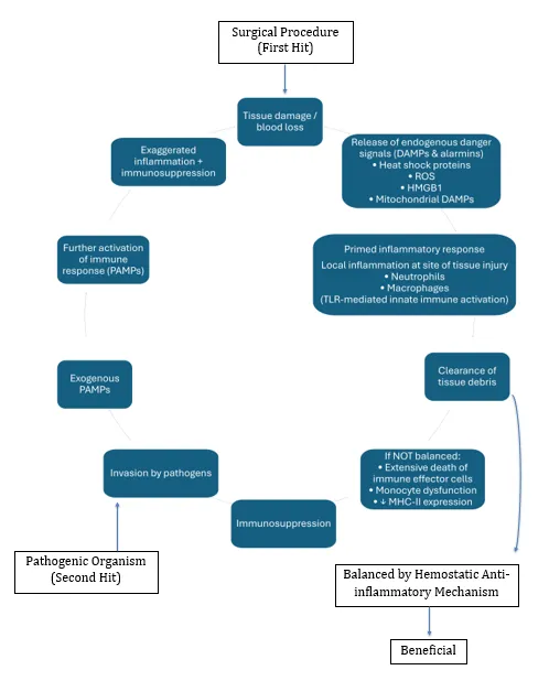
The pathogenesis of SSI can be explained by the combined effects of surgical trauma and pathogenic microbial invasion, which together create a self-perpetuating vicious cycle leading to infection. Surgical intervention constitutes a first hit, producing tissue injury and blood loss that trigger the release of damage-associated molecular patterns (DAMPs), including heat shock proteins, reactive oxygen species, HMGB1, and mitochondrial components. These signals activate innate immunity through Toll-like receptor–dependent pathways, resulting in a primed inflammatory response. Neutrophils and macrophages are recruited to the site of injury to remove cellular debris and support tissue repair. When this response is effectively regulated by homeostatic and anti-inflammatory mechanisms, healing ensues.
Failure of regulation leads to excessive inflammation, immune cell depletion, monocyte dysfunction, and reduced MHC-II expression, causing postoperative immunosuppression. Subsequent microbial invasion represents a second hit, during which pathogen-associated molecular patterns (PAMPs) interact with DAMPs, amplifying inflammation and immune dysregulation. This results in increased susceptibility to infection and thereby perpetuating a vicious cycle of surgical site infection. This immunological imbalance explains the increased susceptibility to surgical site infection despite adherence to sterile surgical technique. [10] (Figure 1)
6. Etiological Agents
Most SSIs originate from the patient’s endogenous microbiota, primarily derived from the skin, gastrointestinal tract, or mucosal surfaces. These microorganisms gain access to the surgical wound either intraoperatively or during the immediate postoperative period. Table 3 summarizes the spectrum of pathogens associated with surgical site infections (SSIs) in neonatal and pediatric patients.
| Pathogen | Typical Pathogen | Source / Notes |
|---|---|---|
| Category | ||
| Gram-positive cocci | Staphylococcus aureus (MSSA/MRSA) | S. aureus is the most frequently detected pathogen in superficial SSIs in neonates and pediatric surgical wounds. It is also commonly implicated in orthopedic, neurosurgical, and cardiac surgeries. |
| Coagulase- negative staphylococci (CoNS) | CoNS (e.g., Staphylococcus epidermidis) are often the second most common cause of SSIs, particularly in neurosurgical procedures. Their ability to form biofilms on implants and devices contributes to persistent infection. |
Enterococcus spp. Normal gastrointestinal commensals that are frequently implicated in SSIs, especially following abdominal, colorectal, and urological procedures.
| Gram-negative bacilli | Escherichia coli | Escherichia coli is among the most frequently isolated pathogens in deep and organ-space SSIs. It is the most common organism (20–40%) isolated in urological surgical site infections. |
|---|---|---|
| Klebsiella pneumoniae | Characterized by a prominent polysaccharide capsule, which enhances virulence and immune evasion. Increasingly associated with multidrug- resistant and ESBL-producing strains in neonatal and pediatric SSIs. | |
| Pseudomonas aeruginosa | Commonly implicated in SSIs among neonates and children with burns, prolonged ICU/NICU stay, or prior antibiotic exposure. Produces the | |
| characteristic blue-green pigment (pyocyanin) and forms biofilms. | ||
| Acinetobacter baumannii | Identified in wound and nosocomial surgical infections. Considered an emerging threat in NICUs, particularly due to multidrug resistance. | |
| Anaerobes | Bacteroides spp. | Isolated from neonatal abdominal SSIs, including post-necrotizing enterocolitis (NEC) surgery and bowel perforation repairs. Less frequently reported in neonates than in older children. |
Fungal Candida spp. Cause of hospital-acquired SSIs, particularly in children with prolonged antibiotic therapy, central venous lines, total parenteral nutrition (TPN), or immunocompromised states.
Polymicrobial Mixed aerobic and Commonly observed following gastrointestinal anaerobic flora or bowel procedures, contaminated or dirty surgeries, and in cases of perforation or peritonitis.
7. Clinical Features
Superficial incisional: pain, erythema, warmth, swelling, tenderness, purulent discharge from skin or subcutaneous tissue; usually minimal systemic signs.
Deep incisional SSI: Severe pain, wound induration, deep purulent discharge, wound separation or dehiscence, fever, abscess formation.
Organ/space SSI: Fever, sepsis, localized or generalized abdominal pain, ileus, anastomotic leak, peritonitis, abscess on imaging, organ dysfunction.
In neonates, systemic signs may be subtle or absent, with feeding intolerance or temperature instability often being the earliest indicators.
8. Diagnosis
Diagnosis is based on clinical findings and microbiological evaluation of wound discharge, supported by laboratory investigations such as elevated white blood cell count, C-reactive protein, and procalcitonin, as well as imaging wherever indicated. Superficial surgical site infections are primarily diagnosed clinically, whereas deep and organ/space infections generally require imaging, with ultrasonography preferred in children and computed tomography reserved for complex cases.
9. Prevention Of Surgical Site Infections [13, 14]
9.1. Pre-operative Bathing-A shower or bath using plain soap or antiseptic soap should be performed on the day of surgery or the day before. Chlorhexidine has not demonstrated any added advantage in reducing SSI rates in pediatric patients over plain soap. Therefore, routine chlorhexidine bathing is not recommended.
9.2. Hair Removal-Routine pre-operative hair removal is not recommended. Hair removal is necessary to facilitate the surgical procedure and should ideally be performed on the day of surgery using electric clippers should be used. Razors should be avoided due to the risk of micro-abrasions and increased SSI risk.
9.3. Patient Attire-Clean, procedure-appropriate theatre clothing should be provided to the patients in order to facilitate access to the operative site while maintaining dignity.
9.4. Surgical Hand Antisepsis-Surgical hand washing is among the most critical factors affecting the risk of SSIs, which result in a considerable burden of morbidity and mortality. Hand rubbing and hand scrubbing have not demonstrated superiority of one over the other in reducing surgical site infection rates when performed with appropriate technique. [Figure 2] [Table 4] Povidone-iodine, chlorhexidine gluconate and alcohol-based surgical scrub are among the various Surgical hand scrubs utilized. Hands and nails must be visibly clean, and single-use nail cleaners are recommended.
| Feature | Surgical Hand Scrubbing | Surgical Hand Rubbing |
|---|---|---|
| Method | Hands and forearms washed with water + antiseptic using a scrub/brush | Alcohol-based hand rub applied without water |
| Agents used | Chlorhexidine (2–4%), povidone-iodine (4–7.5%) | Alcohol (60 –85%) ± chlorhexidine |
| Water required | Yes | No |
| Duration | 3–5 minutes | 1.5–3 minutes |
| Mechanical action | Mechanical friction (brush/sponge) | Chemical action only |
| CFU reduction | Good | Equal or better immediate CFU reduction |
| Skin tolerance | More skin irritation | Better skin tolerance |
| Agent | Common Concentration | Residual Effect | Advantages | Disadvantages |
|---|---|---|---|---|
| Chlorhexidine gluconate (CHG) | 2%–4% | Yes (up to 6 h) | Persistent action, good skin adherence | Skin irritation, rare anaphylaxis |
| Povidone-iodine (PVP-I) | 4%–7.5% | No | Broad spectrum, inexpensive | No residual effect, skin staining |
| Alcohol-based scrubs (ethanol / isopropanol / n- propanol) | 60–85% | No (unless combined with CHG) | Rapid action, best Colony Forming Unit (CFU) reduction, skin- friendly | No cleaning action |
| Alcohol + CHG combination | Alcohol + 0.5–1% CHG | Yes | Rapid + persistent effect | Cost |
| Hexachlorophene (obsolete) | 3% | Yes | Persistent action | Neurotoxicity |
| Triclosan | 1% | Minimal | Previously popular | Resistance concerns |
9.5. Skin Antisepsis at the Surgical Site.
Skin preparation should be performed immediately before incision using: [Table 6]
- Alcohol-based chlorhexidine, or
- Povidone-iodine 10% if chlorhexidine is contraindicated.
The WHO guidelines on the prevention of SSIs recommend chlorhexidine-alcohol rather than aqueous povidone-iodine or povidone-iodine with alcohol for surgical skin preparation.
| Agent | Concentration | Type | Advantages | Limitations / Notes |
|---|---|---|---|---|
| Chlorhexidine + Alcohol | 2% CHG in 70% alcohol | Preferred | Rapid action, residual effect, best SSI reduction | Avoid mucosa, eyes, open wounds |
| Alcohol (ethyl / isopropyl) | 70% | Alternative | Very rapid bactericidal action | No residual effect, flammable |
| Povidone- iodine solution | 10% | Traditional | Broad spectrum | No residual effect, inactivated by organic matter. |
| Povidone- iodine + alcohol | 10% PI + alcohol | Alternative | Better than aqueous iodine | Less persistent than CHG |
| Aqueous chlorhexidine | 2–4% | When alcohol contraindicated | Residual activity | Slower onset |
| Iodine tincture (obsolete) | 2% iodine in alcohol | Historical | Rapid action | Skin irritation |
9.6. Decolonization of nasal carriers of Staphylococcus aureus- Perioperative intranasal mupirocin (2%), applied to both nostrils twice daily for 5 days with or without chlorhexidine body wash, is recommended for selected patients with documented Staphylococcus aureus nasal carriage, prior to surgery. Routine universal decolonization is however, not recommended due to concerns regarding mupirocin resistance
9.7. Maintenance of Normothermia-Peri-operative hypothermia is associated with an increased risk of SSI. Forced-air warming blankets, Warmed intravenous fluids, Maintenance of appropriate operating room temperature should be used.
9.8. Fluid Management-Maintenance of normovolemia is essential. Both hypovolemia and fluid overload should be avoided. Adequate tissue perfusion supports wound healing and immune function.
10. Treatment
The management of surgical site infections has evolved from prolonged empirical antibiotic therapy to a multimodal, evidence-based approach emphasizing timely source control, culture-guided antimicrobial therapy, adjunctive wound care strategies such as negative pressure wound therapy (NPWT), and multidisciplinary management involving surgeons, infectious disease specialists, microbiologists, and wound care nurses. Antimicrobial stewardship is integral to this approach to optimize outcomes and limit the development of resistance. Table 7 summarizes the general principles of management of surgical site infections.
| SSI Type | Management |
|---|---|
| Superficial incisional | Local wound care, drainage if required, and targeted antibiotic |
| SSI | therapy |
| Deep incisional SSI | Surgical drainage ± debridement with systemic antibiotic therapy |
| Organ/space SSI | Surgical intervention with prolonged, culture -guided antimicrobial therapy |
References
- Kamat US, Fereirra AMA, Kulkarni MS, Motghare DD. A prospective study of surgical site infections in a teaching hospital in Goa. Indian J Surg 2008; 70: 120– 124.
- Arora A, Bharadwaj P, Chaturvedi H, Chowbey P, Gupta S, Leaper D, et al. A review of prevention of surgical site infections in Indian hospitals based on global guidelines for the prevention of surgical site infection, 2016. J Patient Saf Infect Control 2018; 6: 1.
- Suri V, Singh H, Mohindra R, Suri A, Prasad D, Bhalla A. Prevention of surgical site infections: Where do we stand today? Curr Med Res Pract 2024; 14: 95.
- Srivastav S, Khurana S, Mukhopadhyay C, Myatra SN, Katyal S, Katoch O, et al.
- Surveillance for surgical site infections developed during hospital stay & after discharge: A multicentric study. Indian J Med Res 2024; 160: 428–437.
- Kamath R, Kumar AS, Bari T, Rj V, Kamath S, Sugunan A, et al. Risk Factors for Surgical Site Infections after Paediatric Appendectomies in a Tertiary Care Teaching Hospital in Coastal Karnataka. Epub ahead of print 6 June 2025. DOI:
- 12688/f1000research.163894.1.
- Catania VD, Boscarelli A, Lauriti G, Morini F, Zani A. Risk Factors for Surgical Site Infection in Neonates: A Systematic Review of the Literature and Meta- Analysis. Front Pediatr 2019; 7: 101.
- Meoli A, Ciavola L, Rahman S, Masetti M, Toschetti T, Morini R, et al. Prevention of Surgical Site Infections in Neonates and Children: Non-Pharmacological Measures of Prevention. Antibiotics 2022; 11: 863.
- Global guidelines for the prevention of surgical site infection. [Second edition].
- Geneva, Switzerland: World Health Organization, 2018.
- Surgical site infection protocol edition 7.1 Definition of SSI surveillance poster, https://www.nss.nhs.scot/media/3469/definitions-of-surgical-site-infections-surveillance.pdf (accessed 26 January 2026).
- Cui P, Fang X. Pathogenesis of infection in surgical patients. Curr Opin Crit Care 2015; 21: 343–350.
- Al Jarousha AMK, El Jadba AHN, Al Afifi AS, El Qouqa IA. Nosocomial multidrug-resistant Acinetobacter baumannii in the neonatal intensive care unit in Gaza City, Palestine. Int J Infect Dis IJID Off Publ Int Soc Infect Dis 2009; 13: 623– 628.
- Surgical site infection - etiology, risk factors, and preventive measures – a literature review. Med Sci; 13. Epub ahead of print 4 June 2025. doi:10.53453/ms.2025.6.2.
- Allegranzi B, Bischoff P, De Jonge S, Kubilay NZ, Zayed B, Gomes SM, et al. New WHO recommendations on preoperative measures for surgical site infection prevention: an evidence-based global perspective. Lancet Infect Dis 2016; 16: e276–e287.
- Meoli A, Ciavola L, Rahman S, Masetti M, Toschetti T, Morini R, et al. Prevention of Surgical Site Infections in Neonates and Children: Non-Pharmacological Measures of Prevention. Antibiotics 2022; 11: 863.