
I. Introduction:
Vascular anomalies are heterogenous group of anomalies seen in pediatric age group involving abnormal development of blood vessels and lymphatic channels. Most of them are noted at birth or during infancy. For centuries, they have been referred by various overlapping clinical and histopathologic terms which led to misdiagnosis and inappropriate management1. In the nineteenth century, Virchow categorized the vascular anomalies on the basis of histopathologic features2. The ISSVA classification was first developed by Mulliken and Glowacki about 30 years ago based on clinical behaviour, endothelial characteristics, and natural history and is now the universally accepted classification for vascular anomalies.3,4,5
II. Classification
The ISSVA Classification broadly divides the vascular anomalies into vascular tumours and vascular malformations.5 Vascular tumours are characterized by endothelial hyperplasia; vascular malformations arise due to vascular dysmorphogenesis and exhibit normal endothelial turnover.5 Table 1 depicts the classification in simple tabular form.
| Category | Subclassification | Lesions included | ||
|---|---|---|---|---|
| 1 | Vascular Tumours | Benign Rare Benign and Intermediate Grade Vascular Tumours Intermediate and Borderline Malignant Vascular Tumours Malignant Vascular Tumours | Infantile Hemangioma Congenital hemangioma (RICH, NICH, PICH) Hepatic Hemangioma Kaposiform Hemangioendothelioma (KHE) Pyogenic Granuloma Tufted Spindle Cell Epithelioid Haemangioma Epithelioid haemangioendothelioma Angiosarcoma Hepatic Angiosarcoma Kaposi Sarcoma | |
| 2 | Vascular Malformations | Simple malformations | Slow flow Malformations High flow Malformations Capillary-venous (CV) | Capillary malformation (port- wine stain) Venous malformation Lymphatic malformation (macro-/microcystic) Arteriovenous malformation (AVM) Arteriovenous Fistula |
| Combined malformations Malformations associated with syndromes | Capillary-lymphatic (CL) Capillary-lymphatic Klippel–Trénaunay syndrome Parkes Weber syndrome Sturge–Weber syndrome CLOVES syndrome | -venous (CLV) |
Table 1: ISSVA Classification of Vascular Anomalies
Accurate classification of these anomalies helps to guide treatment decisions (observation, oral medications, sclerotherapy, surgery and/or embolization) in neonatal period itself and identify lesions associated with life-threatening complications like cardiac failure or Kasabach–Merritt phenomenon.3,6 This facilitates parental counselling, prognostication, management and follow-up.
III. Diagnosis
Radiology plays a central role in diagnosis, classification, treatment planning, monitoring response to sclerotherapy or embolization, detecting recurrence or progression and assessing complications (thrombosis or tissue ischemia) and follow-up of vascular malformations.
Ultrasonography (USG) and Doppler: Ultrasound is non-invasive, readily available, and does not involve ionizing radiation.7 It helps to delineate the morphology, echogenicity, volume, extent, type of the cystic spaces (if present) and the presence of phleboliths.
- Venous malformations can be seen as hypoechoic or heterogeneous compressible lesions; may contain phleboliths (echogenic foci with shadowing)8
- Lymphatic malformations mostly comprise of multiloculated cystic lesions -
macrocystic, microcystic, or mixed4
- Arteriovenous malformations (AVMs) are ill-defined heterogeneous masses without discrete cysts7
Doppler ultrasound differentiates low-flow lesions (e.g., capillary, venous, lymphatic) which show absent or minimal flow and high-flow lesions (e.g., AVMs, AV fistulas) which show high-velocity arterial flow, low-resistance waveforms, arteriovenous shunting.9
Magnetic Resonance Imaging (MRI): MRI is the gold standard for comprehensive evaluation of vascular malformations and also for follow-up of lesions.8,10 It provides superior soft-tissue contrast and accurate assessment of the extent of the lesion. The MRI characteristics of the various vascular malformations are as follow:
- Venous malformations8,10:
- T1-weighted: Hypointense to isointense
- T2-weighted: Markedly hyperintense
- Phleboliths appear as signal voids.
- Lymphatic malformations11:
- Multicystic lesions with fluid–fluid levels
- T2 hyperintensity; minimal enhancement
- Arteriovenous malformations11:
- Flow voids on T1 and T2
- Enlarged feeding arteries and draining veins
Dynamic contrast-enhanced MRI allows differentiation of low-flow vs high-flow lesions, mapping of feeding arteries and draining veins and pre-interventional planning.10,11,12
Computed Tomography (CT): CT has a limited role due to ionizing radiation but is useful in selected situations like detection of phleboliths in venous malformations, evaluation of bony involvement and emergency assessment of bleeding or airway compromise.13 CT angiography demonstrates high-flow lesions but is avoided in neonates unless MRI is contraindicated.13
Conventional Angiography (Digital Subtraction Angiography – DSA): DSA remains the gold standard for high-flow vascular malformations for planning interventions.11,14 Indications include precise delineation of arteriovenous shunts, identification of feeding vessels and therapeutic embolization during the same session.11,14
IV. Vascular Tumours
Vascular tumours are heterogeneous group of lesions ranging from benign, self-limited proliferations to highly aggressive malignant neoplasms. While infantile haemangioma and related benign tumours account for the majority of vascular tumours in childhood, a small subset of rare and malignant vascular tumours is clinically significant because of their aggressive behaviour, diagnostic difficulty, and therapeutic challenges.
1. Benign Vascular Tumours
A) Infantile Haemangioma
Introduction and Epidemiology: Infantile haemangioma (IH) is the most common benign vascular tumour of infancy seen in approximately 4–10% of infants.15,16,17 They are true vascular neoplasms arising from endothelial cell proliferation and may be absent or only subtly evident at birth.15,16,17 There is a marked female predominance, with a female-to-male ratio ranging from 3:1 to 5:1. Risk factors include multiple gestations, advanced maternal age, placental abnormalities, Caucasian ethnicity, prematurity and low birth weight. Most lesions become clinically apparent within the first 2–4 weeks of life.16,17
Natural History: Infantile haemangiomas follow a predictable natural history - triphasic course comprising three phases: proliferative phase, plateau phase, and involution phase. Rapid growth usually occurs during the first 3– months, with proliferation occasionally continuing up to 9–12 months. Involution begins after the first year and progresses gradually over several years. Approximately 50% involute by years, 70% by 7 years, and up to 90% by 9–10 years. Residual skin changes may persist despite complete involution.16,17 Etiopathogenesis: The etiopathogenesis of infantile haemangioma is multifactorial. The placental origin theory is supported by the expression of glucose transporter protein-1 (GLUT- 1) in infantile haemangiomas.17 Hypoxia-driven angiogenesis represents another mechanism. Perinatal hypoxic stress leads to upregulation of hypoxia-inducible factor-1α (HIF-1α), which stimulates angiogenic mediators such as vascular endothelial growth factor (VEGF) and basic fibroblast growth factor (bFGF). This hypothesis explains the increased incidence of infantile haemangiomas in premature and low-birth-weight infants.17 The renin–angiotensin system has also been implicated. Elevated renin levels in early infancy might promote angiogenesis through angiotensin II–mediated endothelial proliferation, providing an explanation for the dramatic therapeutic efficacy of beta-adrenergic blockers, particularly propranolol.15,17
Classification: Infantile haemangiomas can be classified according to depth into superficial, deep, or mixed; and as per morphology and distribution into focal, segmental, multifocal, or indeterminate. Segmental haemangiomas are typically larger, plaque-like lesions following embryological or developmental segments and are associated with a higher risk of complications and syndromic associations.17,18
Clinical Features: Clinically, these are soft, compressible lesions that blanch on pressure. Superficial lesions appear as bright red, well-demarcated plaques, whereas deep lesions present as bluish subcutaneous swellings beneath normal skin. Most lesions are asymptomatic, although rapid growth may predispose to complications.16 Head and neck region is most commonly affected, accounting for nearly 60% of cases, followed by the trunk and extremities (Figure 1). Visceral haemangiomas are uncommon, with the liver being the most frequently involved internal organ. The presence of multiple cutaneous haemangiomas should raise suspicion of visceral involvement.16,17
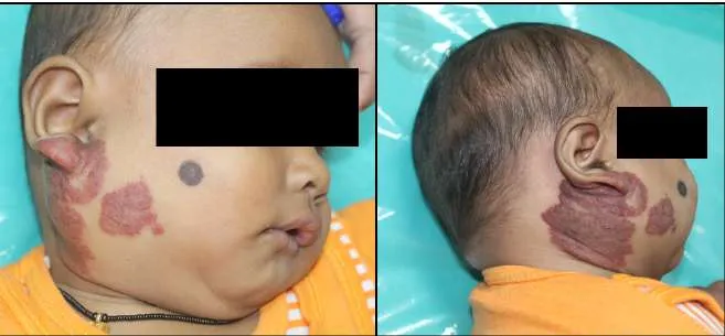
Complications: Complications occur in approximately 10–15% of cases. Ulceration is the most common complication and may lead to pain, bleeding, infection, and scarring. Lesions in critical locations may cause functional impairment, including visual obstruction, airway compromise, feeding difficulties, or hearing impairment.16,17 Large or multiple haemangiomas may result in high-output cardiac failure. Syndromic Associations: Large segmental facial haemangiomas may be associated with PHACE syndrome, while extensive segmental haemangiomas involving the lower body may be associated with LUMBAR, PELVIS, or SACRAL syndromes, characterized by spinal, genitourinary, and anorectal anomalies.17,18
Investigations: Diagnosis is primarily clinical. Ultrasonography with Doppler is useful for deep or atypical lesions. Magnetic resonance imaging (MRI) is indicated for large, deep, or segmental haemangiomas, particularly when syndromic associations are suspected.
Echocardiography and thyroid function tests should be done in infants with extensive or multiple lesions.17,18
Differential Diagnosis: The differential diagnosis includes congenital haemangiomas, vascular malformations, pyogenic granuloma, Kaposiform Hemangioendothelioma , and other soft tissue tumours.
Management: Most infantile haemangiomas require observation alone. Treatment is indicated for lesions that are life-threatening, ulcerated, rapidly proliferating, functionally impairing, or at risk of permanent disfigurement.18,19
Medical Therapy: Problematic infantile haemangiomas are managed by oral propranolol administered at 2–3 mg/kg/day in divided doses with appropriate monitoring. Treatment is typically continued for 6–12 months, often until 12–1 months of age.15,18,19,20 Topical Timolol is effective for small, superficial cutaneous lesions. Systemic corticosteroids are reserved for rare refractory cases.19
Surgical and Laser Therapy: Pulsed dye laser therapy is useful for ulcerated lesions and residual telangiectasia. Surgical intervention is rarely required during infancy but may be considered for residual deformity or complications unresponsive to medical therapy.17,19
Prognosis: The prognosis of infantile haemangioma is excellent, with most lesions undergoing spontaneous involution. Early identification and timely intervention in high-risk cases significantly reduce morbidity and optimize cosmetic and functional outcomes.17,18
B) Congenital Haemangioma
Introduction and Epidemiology: Congenital haemangioma is uncommon and accounts for less than 1% of all vascular tumours in infancy.21,22 It does not exhibit the characteristic natural history seen in infantile haemangiomas; instead follows a distinct biological and clinical course. The lesions arise from aberrant vascular development during intrauterine life and demonstrate variable patterns of involution.24,25 They are typically solitary and sporadic, with no established hereditary pattern. Most lesions are identified antenatally or immediately at birth.21,23 There is no gender predilection.
Etiopathogenesis: The exact pathogenesis of congenital haemangioma remains uncertain. These lesions are believed to result from abnormal vasculogenesis during fetal development and do not express GLUT-1.21,24,25 Molecular studies suggest dysregulation of pathways involved in vascular maturation and stabilization.22
Classification: Congenital haemangiomas are classified according to their postnatal behaviour into three types:
- Rapidly involuting congenital haemangioma (RICH)
- Non-involuting congenital haemangioma (NICH)
- Partially involuting congenital haemangioma (PICH)
RICH shows substantial regression within the first year of life, often beginning soon after birth. NICH persists without significant involution, while PICH demonstrates partial regression followed by stabilization.21,23,25
Clinical Features: Congenital haemangiomas most commonly involve head and neck, trunk, or extremities. Lesions are typically well-circumscribed, firm, round or oval masses and may be warm, or pulsatile because of high blood flow (Figure 2). The overlying skin may be violaceous or bluish, often with coarse telangiectasia, or a pale peripheral halo, or occasional central ulceration.21,23 In RICH, progressive softening and flattening occur during infancy, often leaving residual skin atrophy or fibrofatty tissue. NICH lesions remain stable, while PICH lesions show intermediate behaviour.21,25 Histologically, these consist of lobules of capillary-sized vessels separated by fibrous stroma. The endothelial cells are flattened.
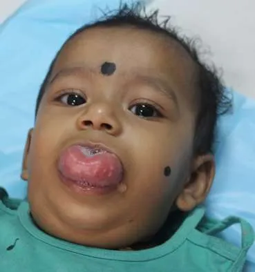
Complications: Ulceration and bleeding are the most common complications. Rarely, large high-flow lesions may result in high-output cardiac failure in the neonatal period. Functional impairment may occur when lesions involve critical anatomical sites.21,23,25
Investigations: Diagnosis is primarily clinical, supported by Ultrasonography with Doppler which demonstrates a well-defined high-flow vascular mass. MRI shows a lobulated lesion with flow voids and intense contrast enhancement, providing excellent anatomical delineation.21,22
Differential Diagnosis: The main differential diagnoses include infantile haemangioma, vascular malformations, Kaposiform Hemangioendothelioma, and arteriovenous malformations. Management: Management depends on subtype, symptoms, and associated complications. RICH is usually managed conservatively with observation due to predictable spontaneous involution. NICH and PICH may require intervention when associated with functional impairment, cosmetic concern, ulceration, or bleeding.21,23,25 Medical therapies (beta-blockers and corticosteroids) is generally ineffective. Surgical excision is the treatment of choice for symptomatic non-involuting lesions. Embolization may be used as an adjunct in selected high-flow lesions.22,23
Prognosis: Prognosis varies according to subtype. RICH has an excellent prognosis, though residual skin changes may persist. NICH and PICH may remain stable or require surgical management but never undergo malignant transformation.21,23,25
C) Hepatic Hemangioma
Introduction and Epidemiology: Hepatic hemangioma is rare but represents the most frequent hepatic vascular tumour in infancy.26,27 It represents a spectrum of vascular neoplasms arising from endothelial proliferation within the hepatic parenchyma and is commonly diagnosed in infants with multiple cutaneous hemangiomas.27,28 There is a female predominance; Prematurity and low birth weight are recognized risk factors.28
Classification: Hepatic hemangiomas are broadly classified based on number and distribution of lesions into focal, multifocal, and diffuse types.27 Focal hepatic hemangioma is usually a solitary lesion, often present at birth. Multifocal hepatic hemangioma consists of multiple discrete lesions scattered throughout the liver parenchyma and is typically associated with cutaneous infantile hemangiomas.26 Diffuse hepatic hemangioma is characterized by near-total replacement of hepatic parenchyma by innumerable hemangiomatous lesions and represents the most severe form of the disease.27
Etiopathogenesis: These arise from dysregulated angiogenesis driven by hypoxia-induced pathways and overexpression of angiogenic factors such as vascular endothelial growth factor.24 Endothelial cells in infantile-type hepatic hemangiomas express GLUT-1.29 In diffuse and multifocal forms, extensive arteriovenous shunting within the liver leads to significant hemodynamic consequences.30 Hepatic hemangiomas also express type 3 iodothyronine deiodinase, which accelerates degradation of thyroid hormones and may result in consumptive hypothyroidism.31,32 Histologically, infantile-type hepatic hemangiomas are composed of lobules of capillary-sized vessels lined by plump endothelial cells with mitotic activity during the proliferative phase.29
Clinical Features: The clinical presentation of hepatic hemangioma varies widely.27,30 Focal asymptomatic lesions may be detected incidentally on antenatal or postnatal imaging. Multifocal lesions may present with hepatomegaly, abdominal distension, or signs related to associated cutaneous hemangiomas.28 Diffuse hepatic hemangioma presents early in infancy with massive hepatomegaly, abdominal compartment symptoms, and systemic complications.30
Complications: The most serious complication of hepatic hemangioma is high-output cardiac failure, particularly in diffuse disease.30 Consumptive hypothyroidism is another unique complication caused by increased inactivation of thyroid hormones within the tumour tissue.31,32 Other complications include abdominal compartment syndrome, hepatic dysfunction due to parenchymal replacement, and, rarely, hemorrhage.27,30 Affected infants may also develop respiratory distress, failure to thrive , and neurodevelopmental impairment.31,32
Investigations: Ultrasonography with Doppler typically demonstrates well-defined, hyper-vascular lesions with high-flow signals.27,33 On MRI, the lesions are typically hypointense on T1-weighted images and hyperintense on T2-weighted images, with intense, homogeneous enhancement following contrast administration.33 Laboratory evaluation should include thyroid function tests in all infants with multifocal or diffuse hepatic hemangiomas.31,32 Echocardiography is essential to assess cardiac function and detect high-output failure.
Differential Diagnosis: The differential diagnosis includes hepatic arteriovenous malformations, mesenchymal hamartoma, hepatoblastoma, and metastatic neuroblastoma.27
Management: The management depends on clinical presentation and extent of disease.27,30 Asymptomatic focal lesions may be managed conservatively with observation and serial imaging. Oral propranolol is the first-line therapy for reduction in tumour size, improvement in cardiac function, and resolution of hypothyroidism when combined with appropriate thyroid hormone replacement.6,31,34 Corticosteroids, interferon-α, and vincristine are reserved for refractory cases.27,34 Interventional radiology procedures such as hepatic artery embolization may be considered in severe, treatment-resistant cases.30 Surgical resection and liver transplantation may be required in select infants with uncontrollable heart failure or hepatic failure.30
Prognosis: The prognosis depends on disease extent and timeliness of treatment.27,30 Focal lesions have excellent prognosis.30 Multifocal and diffuse hepatic hemangiomas, now have significantly improved outcomes with early diagnosis and propranolol therapy.6,34 Long-term follow-up is essential to monitor for residual hepatic changes and neurodevelopmental outcomes, particularly in infants with previous hypothyroidism or cardiac failure.31,32
D) Kaposiform Haemangioendothelioma
Definition and Epidemiology: Kaposiform haemangioendothelioma (KHE) is a rare vascular neoplasm which arises from endothelial cells, typically presenting in neonatal period or early infancy.35 It is characterized by infiltrative growth into skin, subcutaneous tissue, muscle, and occasionally deeper structures such as bone or retroperitoneum.35,36 Although benign, KHE behaves in a locally aggressive manner and does not undergo spontaneous involution.6 The majority of lesions are sporadic, with no clear genetic predisposition.35
Etiopathogenesis: The exact pathogenesis of KHE is not fully understood.35 The tumour is believed to arise from dysregulated angiogenesis and lymphangiogenesis during development.24 It does not express GLUT-1.37 Molecular studies suggest involvement of signalling pathways related to vascular proliferation and platelet trapping, which underlies its association with Kasabach–Merritt phenomenon.38
Clinical Features: It presents as an ill-defined, firm, indurated, violaceous or purpuric mass that infiltrates surrounding tissues, often associated with pain, swelling, and local warmth.6,35 Lesions commonly involve the extremities, trunk, retroperitoneum, and cervicofacial region and show progressive enlargement.6,35,36 Deep lesions may present with nonspecific symptoms related to mass effect, while cutaneous lesions may show rapid enlargement accompanied by bruising, suggesting the onset of Kasabach–Merritt phenomenon.3,38
Histologically, KHE is characterized by nodules of spindle-shaped endothelial cells forming slit-like vascular channels, interspersed with areas of lymphatic differentiation, resembling Kaposi sarcoma but lacking malignant features.35,36 Immunohistochemistry typically shows positivity for CD31 and CD34.37
Kasabach–Merritt Phenomenon: Kasabach–Merritt phenomenon is a severe consumptive coagulopathy characterized by thrombocytopenia, elevated fibrin degradation products, hypofibrinogenemia, and anaemia due to platelet trapping and consumption within a vascular tumour.3,38 It occurs almost exclusively in association with KHE and tufted angioma.6,38 The pathophysiology of KMP involves sequestration and activation of platelets within the abnormal vascular channels of the tumour which leads to platelet aggregation, fibrin deposition, and secondary activation of the coagulation cascade, resulting in consumption of clotting factors.38 Progressive intratumoral thrombosis causes further tumour enlargement, creating a vicious cycle of growth and coagulopathy.3 Figure 3 shows an infant with KMP.
Clinically, KMP presents with sudden enlargement of the vascular tumour, overlying purpura or ecchymosis, and systemic signs of bleeding such as petechiae, mucosal haemorrhage, or gastrointestinal bleeding.3,38 Affected infants may appear acutely ill and are at high risk of life-threatening hemorrhage.3,39 Laboratory evaluation reveals severe thrombocytopenia, often with platelet counts below 20,000/mm³. Fibrinogen levels are markedly reduced, and D-dimer levels are elevated.38 There is prolongation of prothrombin time and activated partial thromboplastin time.39
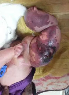
Investigations: Ultrasonography with Doppler demonstrates a heterogeneous, infiltrative soft-tissue mass with variable vascularity.7 Magnetic resonance imaging is the modality of choice for defining tumour extent and typically shows a poorly circumscribed lesion with intermediate to low signal intensity on T1-weighted images and heterogeneous high signal intensity on T2-weighted images, with infiltrative margins.7,27 Management: The management of KHE complicated by KMP is challenging and requires a multidisciplinary approach involving haematology, oncology, surgery, radiology and intensive care.3,38 Supportive care includes careful monitoring, transfusion of blood products for life-threatening bleeding, and avoidance of platelet transfusions unless there is active haemorrhage, as transfused platelets may worsen tumour growth.3,38
Medical therapy: Sirolimus, an mTOR inhibitor, has emerged as first-line therapy due to its efficacy in controlling tumour growth and resolving coagulopathy.40,41,42 Sirolimus inhibits the PI3K–AKT–mTOR pathway, reducing endothelial proliferation, platelet trapping, and angiogenic signaling.40 Corticosteroids and Vincristine may still play a role in combination therapy or in refractory cases.6,35,39
Alpelisib (PI3K-α Inhibitor) is an emerging targeted therapy which has gained attention as a precision therapy for PI3K-pathway–driven vascular anomalies. It is effective in PIK3CA- or TEK-mutated vascular malformations resistant to Sirolimus.43,44 It has demonstrated radiologic volume reduction, symptom improvement, and D-dimer normalization in selected patients. Side effects like hyperglycaemia, gastrointestinal toxicity, and limited pediatric safety data currently restrict its routine use as first-line therapy.44,45
Surgery: Surgical excision may be considered in localized lesions after medical stabilization.1,4 Embolization and radiotherapy are reserved for selected refractory cases.6,39
Prognosis: The prognosis of KHE depends largely on the presence and severity of KMP.3,38 With advances in medical therapy, particularly the use of sirolimus, survival has improved significantly.40,41,42 However, long-term morbidity related to local tissue destruction, functional impairment, and recurrence may occur, necessitating prolonged follow-up.6,38
E) Pyogenic Granuloma
Introduction and Epidemiology: Pyogenic granuloma, also known as lobular capillary haemangioma, is a common benign vascular lesion characterized by rapid growth and a marked tendency to bleed.46 It accounts for a significant proportion of acquired vascular lesions of the skin and mucous membranes.47 Despite its name, the lesion is neither pyogenic nor granulomatous.46,47 It represents a reactive vascular proliferation that occurs in response to minor trauma, chronic irritation, or hormonal influences.47,48 Pyogenic granuloma can occur at any age but is particularly common in children, adolescents, and pregnant women.47,48 It occurs equally in males and females in childhood.42 Common sites include the skin of the head and neck, upper extremities, oral mucosa, gingiva, lips, and nail folds.47,48 Oral lesions are especially frequent in children and pregnant women.48
Etiopathogenesis: The pathogenesis of pyogenic granuloma is multifactorial and incompletely understood.46,47,48 The lesion is believed to represent an exaggerated angiogenic response to local stimuli such as trauma, irritation, or inflammation.47,48 Increased expression of angiogenic factors (vascular endothelial growth factor and basic fibroblast growth factor) has been demonstrated, leading to rapid capillary proliferation.46,48 They are not true neoplasms and do not exhibit a proliferative–involutional cycle.46,48
Clinical Features: Clinically, pyogenic granuloma appears as a rapidly growing, painless vascular nodule that develops over weeks.47 The lesion is small, red to violaceous, polypoid or pedunculated lesion with a smooth or lobulated surface.46,47 The lesion is friable and bleeds easily on minimal trauma. Ulceration and crusting are common.47 The lesion is typically bright red in early stages and may darken over time.47 Bleeding is a prominent feature and often the presenting complaint.47
Histologically, pyogenic granuloma is characterized by lobules of capillary-sized vessels arranged in a lobular architecture within an edematous stroma.46,47 The capillaries are lined by flattened endothelial cells and separated by fibromyxoid tissue.46 A variable inflammatory infiltrate is present, particularly in ulcerated lesions.48 Immunohistochemical staining is positive for endothelial markers such as CD31 and CD34.46,47
Differential Diagnosis: The differential diagnosis includes infantile haemangioma, congenital haemangioma, amelanotic melanoma, Kaposi sarcoma, bacillary angiomatosis, and vascular malformations.46,47,48 The absence of a postnatal proliferative phase and the characteristic lobular histology aid in diagnosis.46,47
Investigations: The diagnosis of pyogenic granuloma is primarily clinical.47,48 Imaging is rarely required unless the lesion is atypical or deep.48 Histopathological examination following excision is recommended to confirm the diagnosis and exclude malignant mimics.46,47
Management: Complete surgical excision with cauterization of the base is the treatment of choice and allows histological confirmation.46,47 Other treatment modalities include curettage with electrocautery, laser therapy, cryotherapy, and topical or intralesional agents such as beta-blockers or imiquimod in selected cases.48
Prognosis: The prognosis of pyogenic granuloma is excellent with good cosmetic outcomes.46,47,48 The lesion is benign and does not undergo malignant transformation.46 Recurrence rates range from 5% to 15%, depending on treatment modality and completeness of excision.47
2) Rare Benign and Intermediate-Grade Vascular Tumours
A) Tufted Angioma: Tufted angioma is a rare, benign but locally infiltrative vascular tumour presenting in infancy or early childhood.35,49 Clinically, it appears as a slowly enlarging erythematous or violaceous plaque or nodule, most commonly involving the neck, upper trunk, or proximal extremities.35 Histologically, it is characterized by discrete “tufts” or cannonball-like lobules of capillaries scattered throughout the dermis and subcutaneous tissue.35 Although indolent, tufted angioma is closely related to Kaposiform Haemangioendothelioma and may be complicated by Kasabach–Merritt phenomenon¹. GLUT-1 expression is absent.49
B) Spindle Cell Haemangioma: Spindle cell haemangioma is a rare benign vascular tumour that typically presents as a slow-growing subcutaneous mass in the distal extremities of young individuals.35,38 It may occur as a solitary lesion or as multiple nodules, particularly in association with vascular malformation syndromes.35,38 Histologically, the tumour consists of cavernous vascular spaces interspersed with spindle-shaped endothelial cells.35,38 The clinical course is usually benign, although local recurrence is common after excision.35,38
C) Epithelioid Haemangioma: Epithelioid haemangioma is a benign vascular neoplasm characterized by epithelioid endothelial cells and prominent inflammatory infiltrates rich in eosinophils.35,38 It most commonly affects the skin and subcutaneous tissue of head and neck but may also involve bone.35,38 Clinically, lesions present as small, firm nodules that may be painful or pruritic.35,38 Despite its benign nature, epithelioid haemangioma can mimic malignant vascular tumours histologically, necessitating careful pathological evaluation.35,38
3) Intermediate And Borderline Malignant Vascular Tumours
Epithelioid Hemangioendothelioma: Epithelioid haemangioendothelioma is a rare vascular tumour of intermediate malignancy that can arise in soft tissue, liver, lung, and bone.38 It affects both children and adults.3 Histologically, it is composed of epithelioid endothelial cells arranged in cords and nests within a myxohyaline stroma.38 The clinical behaviour is unpredictable, ranging from indolent to aggressive with metastatic potential.38 Complete surgical excision is the preferred treatment when feasible.38
4) Malignant Vascular Tumours
A) Angiosarcoma: Angiosarcoma is a highly malignant vascular tumour arising from endothelial cells and represents the most aggressive neoplasm in this group.35,38 It is extremely rare in children but may occur in association with chronic lymphoedema, prior radiation therapy, or certain genetic syndromes.35,38 It presents as a rapidly enlarging, bruised or purpuric mass that may ulcerate or bleed.35,38 Histologically, it is characterized by atypical endothelial cells forming irregular, anastomosing vascular channels with marked cellular atypia and high mitotic activity.35,38 Prognosis is poor, with a high rate of local recurrence and distant metastasis.35,38
B) Hepatic Angiosarcoma: Hepatic angiosarcoma is an exceptionally rare but highly aggressive malignant vascular tumour of the liver.35,38 It presents with hepatomegaly, abdominal pain, jaundice, or hepatic failure.35,38 Imaging often reveals multifocal haemorrhagic liver masses.35,38 The prognosis is dismal, with limited treatment options, typically involving a combination of surgery and chemotherapy; outcomes remain poor.35,38
C) Kaposi Sarcoma: Kaposi sarcoma is a malignant vascular neoplasm associated with human herpesvirus-8 (HHV-8) seen in immunodeficient children.35,38 It presents as violaceous patches, plaques, or nodules involving the skin, mucosa, and visceral organs.35,38 Histologically, it shows spindle cell proliferation with slit-like vascular spaces.35,38 Management focuses on treatment of the underlying immunodeficiency and systemic therapy for advanced disease.35,38
V. Vascular Malformations
Introduction
Vascular malformations are a heterogeneous group of congenital anomalies resulting from errors in vascular development during embryogenesis3. Unlike vascular tumours, vascular malformations are not characterized by endothelial proliferation; instead, they represent structural abnormalities of blood vessels that are present at birth, although they may not become clinically apparent until later in life.3,36 These lesions grow proportionately with the child, do not involute spontaneously, and often progress over time, leading to functional impairment, cosmetic deformity, and potentially life-threatening complications.36,50
Embryology of the Vascular System
Normal Vascular Development
The vascular system begins to develop during the third week of gestation through the process of vasculogenesis.36,50 Mesodermal cells differentiate into angioblasts, which aggregate to form primitive vascular channels known as blood islands. These channels subsequently coalesce to form a primary capillary plexus.36
Angiogenesis follows vasculogenesis and involves the sprouting, remodelling, and maturation of this primitive plexus into a hierarchical system of arteries, capillaries, veins, and lymphatic vessels.50 During normal embryogenesis (weeks 4–8), arteries and veins differentiate from the primitive capillary plexus under control of molecular pathways including Notch signalling, VEGF, EphrinB2–EphB4, and TGF-β.3,5,36 Formation of a functional capillary network is essential to regulate blood flow and tissue perfusion.3,5,36
Between the fourth and eighth weeks of gestation, selective regression, differentiation, and maturation of the primitive vascular channels occur.
Embryologic Basis of Vascular Malformations
Failure or aberration at any stage of embryogenesis of vascular channels results in persistent embryonic vessels or abnormal vascular connections, resulting in vascular malformations.3,5,36 The timing of the developmental error determines the type of malformation. Early embryologic insults tend to result in complex, combined malformations, whereas later insults produce more localized and simpler lesions.36
Failure of normal capillary differentiation can lead to capillary malformations. Abnormal maturation of venous channels leads to venous malformations, while disruption of lymphatic vessel development results in lymphatic malformations.8,36 Somatic activating mutations in the TEK (TIE2) gene and PIK3CA gene have been identified in many sporadic venous malformations, linking them to the PIK3CA-related overgrowth spectrum (PROS).3,5,36 Persistence of embryonic arteriovenous connections without interposed capillaries results in arteriovenous malformations.3,5,36 The absence of capillaries leads to direct high-pressure arterial flow into the venous system, causing progressive venous dilation, vessel wall remodelling, and recruitment of collateral vessels over time.3,5,36 Genetic studies have identified germline and somatic mutations associated with AVMs, including RASA1, ENG, ACVRL1, and SMAD4, particularly in syndromic forms such as hereditary haemorrhagic telangiectasia (HHT) and capillary malformation–AVM (CM-AVM) syndrome.3,5,36,50
Normal Development of the Lymphatic System
The lymphatic system develops during the fifth to seventh weeks of gestation.3,36 Lymphatic endothelial cells arise from venous endothelial cells, primarily from the cardinal veins, under the influence of transcription factors such as PROX1, SOX18, and COUP-TFII.3,36 These cells bud from the veins to form primary lymph sacs, which subsequently proliferate and remodel into a complex network of lymphatic vessels3,36
Embryologic Basis of Lymphatic Malformations
Lymphatic malformations arise due to sequestration or failure of developing lymphatic channels to establish normal connections with the venous system.3,36 As a result, isolated lymphatic sacs persist and progressively dilate.3,36 The extent and timing of the embryologic insult determine the size, distribution, and type of lymphatic malformation.3,36 Recent studies have identified somatic activating mutations in the PIK3CA gene in many lymphatic malformations, linking them to the PIK3CA-related overgrowth spectrum (PROS).3,36
1) Simple Vascular Malformations
Simple vascular malformations involve a single type of vessel and include capillary, venous, lymphatic, and arteriovenous malformations.51
A) Capillary Malformations
Introduction: Historically referred to as “port-wine stains,” Capillary Malformations (CMs) are the most common type of vascular malformation and represent congenital abnormalities of the capillary network.3,36 They are characterized by ectatic capillary-sized vessels within the superficial dermis and are present at birth.3,36 They are classified as low-flow vascular malformations and are not associated with endothelial proliferation.3,5
Embryology and Pathogenesis: Capillary malformations arise from abnormalities in the development and maturation of the dermal capillary plexus during early embryogenesis involving defective neural regulation of cutaneous blood vessels, leading to persistent vasodilatation and progressive ectasia of capillaries.3,36
Recent genetic studies have identified somatic activating mutations in the GNAQ and GNA11 resulting in dysregulated intracellular signalling pathways, leading to abnormal vascular tone, vessel enlargement, and failure of normal capillary regression.51 Histologically, capillary malformations consist of dilated capillary-sized vessels located predominantly in the superficial dermis.3,36 The endothelial lining is flat and mature, without mitotic activity or multilayering. The basement membrane may be thickened, and perivascular neural elements are often reduced or absent.36 There is no expression of GLUT-1.3,36 Clinical Features: Capillary malformations are present at birth as flat, well-demarcated macular lesions that range in colour from pale pink to deep red or violaceous.3,36 They typically grow proportionately with the child and do not regress spontaneously.3,36 Over time, lesions may darken, thicken, and develop nodularity due to progressive vascular ectasia and associated soft tissue or bony hypertrophy.5,36 Lesions most commonly involve the head and neck region.3,36 Functional impairment may occur depending on the anatomical site, particularly when involving the eyelids, oral cavity, or extremities.36
Types of Capillary Malformations
1. Nevus Simplex (Salmon Patch): Nevus simplex is the most common and benign form of capillary malformation.3,36 It presents as faint pink macules typically located on the glabella, eyelids (“angel’s kiss”), nape of the neck (“stork bite”), or sacral region.3,36 These lesions are caused by transient capillary dilatation and often fade or disappear within the first few years of life, particularly those on the face.36 Nuchal lesions may persist into adulthood.36
2. Port-Wine Stain (Nevus Flammeus): Port-wine stain is the classic and most clinically significant type of capillary malformation.3,36 It presents at birth as a unilateral or segmental flat red patch that darkens to a deep purple colour over time.36 These port-wine stains persist throughout life and may thicken, develop nodules, and bleed in adulthood.36 Facial port-wine stains, particularly those involving the ophthalmic division of the trigeminal nerve, are associated with Sturge–Weber syndrome, which includes leptomeningeal vascular malformations and ocular complications such as glaucoma.51
3. Capillary Malformation with Soft Tissue and Bony Hypertrophy: These capillary malformations are associated with underlying soft tissue and skeletal overgrowth which reflect more extensive embryologic involvement.3,6 These are often part of complex vascular syndromes like Klippel–Trénaunay syndrome, which is characterized by capillary malformation, venous malformation, and limb overgrowth.3,36 The capillary component typically manifests as a geographic port-wine stain over the affected limb.36
4. Capillary Malformation –Arteriovenous Malformation (CM-AVM): Capillary malformation–arteriovenous malformation is a distinct entity associated with germline mutations in the RASA1 or EPHB4 genes and presents with multiple small, round or oval capillary malformations, often with a pale halo.50 These patients are at risk of developing fast-flow lesions, including arteriovenous malformations and arteriovenous fistulas in the skin, brain, or other organs.50 These may lead to serious complications such as haemorrhage or high-output cardiac failure.50
5. Telangiectatic Capillary Malformations: They consist of fine, dilated superficial vessels and may be isolated or part of systemic disorders.5,36 They are seen in conditions such as hereditary haemorrhagic telangiectasia, where capillary and post-capillary venules are abnormal.36 They are prone to bleeding and may involve mucosal surfaces, necessitating careful evaluation for systemic involvement.36 Differential Diagnosis: Capillary malformations are similar to infantile haemangiomas, and typically appear after birth, proliferate rapidly, and later involute.3,36 Other differential diagnoses include erythema simplex, cutis marmorata telangiectatica congenita, and early-stage arteriovenous malformations.36
Imaging and Diagnostic Evaluation: Diagnosis is primarily clinical.3,36 Imaging is indicated when there is suspicion of deeper involvement or associated anomalies.36 Magnetic resonance imaging is useful for assessing soft tissue and central nervous system involvement, particularly in facial capillary malformations.5,36 Doppler ultrasonography helps confirm the low-flow nature of the lesion.36
Management: Treatment is primarily aimed at cosmetic improvement and prevention of complications.5,36 Pulsed dye laser therapy is the treatment of choice and is most effective when initiated early in childhood.5,36 Surgical intervention is rarely required except for nodular or hypertrophic lesions.36 Management of syndromic capillary malformations requires a multidisciplinary approach.5,36
Prognosis: Capillary malformations are lifelong conditions.3,36 While many remain stable, others progress with age. Early diagnosis and intervention can significantly improve cosmetic outcomes and reduce long-term morbidity, particularly in lesions associated with systemic involvement.5,36
B) Lymphatic Malformations
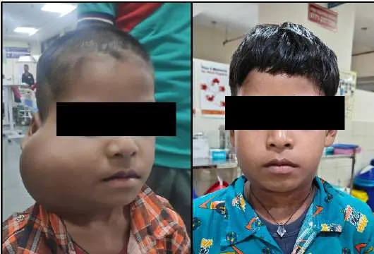
Introduction: Historically termed lymphangiomas, lymphatic malformations (LMs) are congenital, low-flow vascular malformations resulting from abnormal development of the lymphatic system.4,40 They are characterized by dilated lymphatic channels or cystic spaces lined by mature endothelial cells and filled with protein-rich lymphatic fluid.4,40 Lymphatic malformations are present since birth, although they may not become clinically evident until infancy or early childhood.4,40 Figures 4, 5, 6 and 7 show lymphangioma. They neither exhibit endothelial proliferation nor regress spontaneously.4,40
Clinical Features: Lesions are typically soft, compressible, and have transillumination.4,40 Clinical presentation varies depending on lesion size, depth, and anatomical location¹². Complications include infection, haemorrhage, rapid enlargement, airway compromise, and cosmetic deformity.4,40 Histologically, lymphatic malformations are composed of thin-walled, endothelium-lined cystic spaces of varying size, separated by fibrous septa.4,40 The endothelial cells are flat and lack mitotic activity.4,40 The cysts contain clear or straw-coloured proteinaceous fluid and may show haemorrhage or inflammation.4,40 Immunohistochemical staining demonstrates positivity for lymphatic markers such as D2-40 (podoplanin), LYVE-1, and PROX1, confirming lymphatic lineage.4,40
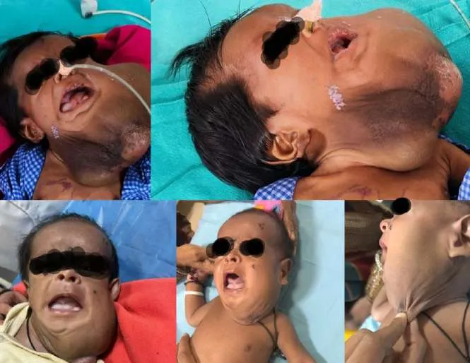
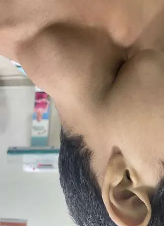
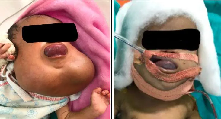
Types of Lymphatic Malformations
1. Macrocystic Lymphatic Malformation: Classically referred to as cystic hygromas, these consist of cysts larger than 2 cm in diameter.4,40 Clinically present as soft, fluctuant, painless swellings that transilluminate brightly; commonly involve the posterior triangle of the neck and axilla.4,40 Ultrasonography demonstrates multiloculated cystic masses; MRI shows well-defined, fluid-filled cysts with septations, hyperintense on T2-weighted images.4,40
2. Microcystic Lymphatic Malformation: Microcystic lymphatic malformations consist of cysts smaller than 2 cm and are often diffuse and infiltrative.4,40 They frequently involve the tongue, lips, floor of the mouth, and skin and present as clusters of small vesicles on skin or mucosa (“frog-spawn” appearance). They are more likely to cause functional impairment such as speech difficulty, feeding problems, and recurrent infections.4,40 Their diffuse nature and infiltrative growth make treatment challenging.4,40
3. Mixed (Macro- and Microcystic) Lymphatic Malformation: Mixed lesions contain both macro- and microcystic components, reflecting earlier and more extensive embryologic involvement.4,41 Combined treatment modalities are often required.4,40
4. Generalized Lymphatic Anomaly: A rare, severe form characterized by multifocal or diffuse involvement of bones, lungs, spleen, and soft tissues.4,40 Patients may present with skeletal destruction, chylous effusions, and respiratory compromise, with significant morbidity and mortality.4,40
5. Gorham–Stout Disease: Also known as vanishing bone disease, it is characterized by progressive osteolysis associated with lymphatic proliferation within bone.4,40 Clinical features include bone pain, pathological fractures, and skeletal deformity.4,40 It is considered part of the spectrum of complex lymphatic anomalies.4,40
Differential Diagnosis: Differential diagnoses include venous malformations, hemangiomas, branchial cleft cysts, thyroglossal duct cysts, and soft tissue tumours.4,40 The absence of postnatal proliferation and low-flow characteristics help distinguish lymphatic malformations from vascular tumours.4,40
Diagnostic Evaluation: Diagnosis is based on clinical findings and imaging studies.4,40 Ultrasonography is useful for initial assessment, while MRI is the imaging modality of choice for delineating lesion extent and involvement of adjacent structures.4,40 Lymphangiography and nuclear medicine studies may be needed in complex cases.4,40
Management: Management is individualized and multidisciplinary.4,40 Treatment options include observation, sclerotherapy, surgical excision, laser therapy, and pharmacologic therapy.4,40
Sclerotherapy: It is the first-line management for macrocystic lesions using bleomycin, doxycycline, or OK-432.4,40 Microcystic lesions may benefit from systemic agents such as sirolimus or laser therapy in refractory cases.4,40
Prognosis: Lymphatic malformations are chronic conditions with variable course.4,40 Many lesions remain stable, while others progress or recur despite treatment.4,40 Early diagnosis and appropriate management improve functional and cosmetic outcomes.4,40
C) Venous Malformations
Introduction: Venous malformations (VMs) represent congenital abnormalities of venous development. They are composed of dysplastic, ectatic venous channels with deficient smooth muscle in the vessel wall.3,36 They are present since birth, although they may not be clinically evident until later in childhood or adolescence.3,36 They do not show endothelial proliferation, do not undergo spontaneous involution, and tend to enlarge progressively with age.3,36 Venous malformations may occur as isolated lesions or as part of complex combined vascular malformations and syndromes.3,36
Clinical Features: Venous malformations present as soft, compressible, bluish lesions that enlarge with dependency, crying, or Valsalva maneuver.3,37 Pain is a common, often due to venous congestion, thrombosis, or localized intravascular coagulopathy.3,36 Lesions may involve skin, subcutaneous tissue, muscle, bone, or visceral organs.3,36 Growth is proportional to somatic growth, but lesions may enlarge rapidly during puberty, pregnancy, trauma, or infection.3,36
Histologically, venous malformations consist of dilated, irregular venous channels lined by flattened, mature endothelial cells.3,376The vessel walls are thin and characteristically deficient in smooth muscle9. Phleboliths, representing organized thrombi, are commonly present.3,36 Immunohistochemistry shows positivity for endothelial markers such as CD31, CD34, and ERG, and negative GLUT-1.3,36
Types of Venous Malformations
1. Simple (Isolated) Venous Malformations: Simple venous malformations involve a single anatomical region and consist exclusively of abnormal venous channels.3,36 They are localized, low-flow, bluish, compressible masses causing pain, swelling, or cosmetic deformity.3,36 Common Sites involved are head and neck, extremities, trunk, oral cavity.3,36 (Figure 7)
2. Infiltrating (Diffuse) Venous Malformations: They extend deeply into muscles, fascia, and bones.3,36 There present with chronic pain, functional impairment, recurrent thrombosis; are poorly circumscribed and difficult to excise.3,36 Complications are Localized intravascular coagulopathy, phlebolith formation, limb dysfunction.3,36
3. Venous Malformations with Phleboliths are commonly seen in long-standing lesions and aid in differentiation on imaging.3,36
4. Glomuvenous Malformations: Glomuvenous malformations (GVMs) are caused by mutations in the GLMN gene.3,36 They present as multiple blue-purple nodules, painful on palpation, less compressible than typical VMs, frequently involve skin.3,36 Histologically, they are composed of venous channels surrounded by glomus cells (modified smooth muscle cells).3,36
5. Familial Cutaneous Venous Malformations: They present as multiple small venous malformations involving skin and mucosa and are inherited in an autosomal dominant pattern, associated with TEK mutations.3,36
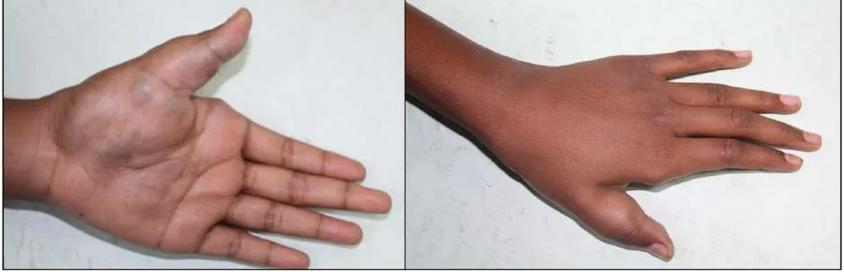
6. Venous Malformations in Syndromic Conditions
- Blue rubber bleb nevus syndrome: Characterized by multiple VMs of skin and GI tract,
chronic bleeding, an aemia.3,36
- Klippel–Trénaunay syndrome: Characterized by capillary malformation, venous
malformation, limb overgrowth.3,36
- Proteus syndrome: Characterized by asymmetric overgrowth with complex vascular
anomalies.3,36
Differential Diagnosis: Differentiation from infantile haemangiomas, lymphatic malformations, arteriovenous malformations, and soft tissue tumours relies on congenital onset, low-flow nature, absence of proliferation, and lack of spontaneous regression.3,36
Diagnostic Evaluation: Ultrasonography with Doppler demonstrates compressible, low-flow venous channels.3,36 MRI is preferred for assessing lesion extent, tissue involvement, and phleboliths.3,36 Conventional angiography is reserved for therapeutic planning.3,36 Laboratory evaluation may reveal elevated D-dimer in extensive lesions due to localized intravascular coagulopathy.3,36
Management: Management is individualized and multidisciplinary.3,36 Options include observation, compression therapy, sclerotherapy, surgical excision, and pharmacologic therapy.3,36 Sclerotherapy: The mainstay of management is intralesional sclerotherapy using bleomycin.3,36
Surgery: Surgery is reserved for localized, well-defined lesions or post-sclerotherapy residuals.3,36
Sirolimus: It is an effective systemic therapy for extensive or refractory lesions.3,8,36
Prognosis: Venous malformations are chronic and progressive.3,36 Complete cure is uncommon, and recurrence is frequent. With appropriate management, most patients achieve good functional and cosmetic outcomes.3,36
D) Arteriovenous Malformations
Introduction: Arteriovenous malformations (AVMs) are high-flow vascular malformations characterized by direct connections between arteries and veins without an intervening capillary bed.3,5,36 This abnormal shunting results in altered hemodynamics, progressive vascular dilation, tissue ischemia, and potential systemic effects such as high-output cardiac failure.3,5,36 AVMs are congenital lesions arising from errors in vascular morphogenesis and are present since birth, although they may remain clinically silent until childhood or adulthood.3,5,36 They do not exhibit endothelial proliferation, do not involute, and demonstrate progressive enlargement over time.3,5,36 These are high-flow lesions characterized by low-resistance shunting, increased arterial inflow, arterialization of draining veins, progressive enlargement of the nidus, risk of rupture, ischemia, and cardiac overload.3,5,36 These features account for the progressive and destructive nature of AVMs.
Histologically, AVMs consist of dysplastic arteries and veins connected through a complex nidus.3,5,36 Vessel walls show abnormal smooth muscle organization, fibrosis, and elastic tissue disruption. There is no endothelial hyperplasia or mitotic activity.3,5,36 Immunohistochemistry confirms endothelial differentiation with positivity for CD31, CD34, ERG, and absence of GLUT-1.3,5,36
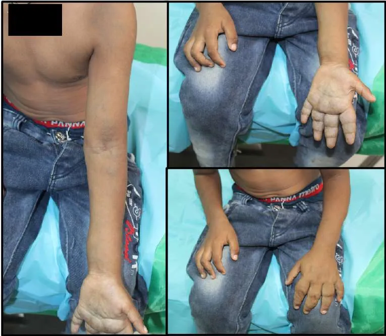
Clinical Features: AVMs may present at any age, often manifesting during hormonal changes, trauma, or infection with features like warm, pulsatile mass, overlying skin erythema, presence of palpable thrill and audible bruit.3,5,36 Complications occur in the form of pain, ulceration, or bleeding, limb overgrowth or ischemia and even high-output cardiac failure in extensive lesions (Figure 8).
Classification and Types
1. Localized (Focal) AVMs: They are confined to a single anatomical region with a discrete nidus and are amenable to targeted embolization or surgery.3,5,36
2. Diffuse AVMs: They are extensive, infiltrative lesions involving multiple tissue planes. They cause progressive tissue destruction, ulceration, bleeding, severe pain, and difficult eradication.3,5,36
3. Arteriovenous Fistulas (AVFs): They are characterised by single direct connection between artery and vein without a nidus; and may be congenital or acquired (trauma/iatrogenic).3,5,36 High-flow AVFs risk cardiac overload; congenital forms may be part of Parkes Weber syndrome.3,5,36
4. Syndromic AVMs
a) CM-AVM syndrome:; They are multiple small capillary malformations and fast-flow lesions affecting skin, brain, lungs, or spine and are associated with RASA1 or EPHB4 mutations.3,36
b) Hereditary haemorrhagic telangiectasia (HHT): It is an autosomal dominant disorder with mucocutaneous telangiectasias and AVMs in lungs, brain, liver, GI tract.3,5,36
5. Visceral AVMs
- Cerebral AVMs: Seizures, headaches, intracranial hemorrhage3,5,36
- Pulmonary AVMs: Right-to-left shunt, hypoxemia, paradoxical emboli3,5,36
- Hepatic AVMs: High-output cardiac failure, portal hypertension3,5,36
Schobinger Clinical Staging3,5,37: AVMs typically progress as follows:
- Stage I (Quiescent): Warmth, blush
- Stage II (Expansion): Pulsation, bruit, pain
- Stage III (Destruction): Ulceration, bleeding, necrosis
- Stage IV (Decompensation): High-output cardiac failure
Differential Diagnosis: AVMs must be distinguished from hemangiomas, venous malformations, lymphatic malformations, and hyper-vascular tumours. Presence of thrill, bruit, and high-flow signals on Doppler strongly favours AVMs.3,5,36
Diagnostic Evaluation: Ultrasonography with doppler confirms high-flow characteristics.3,5,36 MRI helps to define extent and soft tissue involvement.3,5,36 Digital Subtraction Angiography is the gold standard for arterial feeders, nidus, venous drainage, and therapeutic planning.3,5,36
Management: Multidisciplinary approach is required; treatment aims to control symptoms and prevent progression rather than cure.3,5,36 The available options include endovascular embolization, surgical resection, combined embolization and surgery and medical therapy for heart failure in extensive disease. Incomplete treatment may worsen lesion progression due to angiogenic stimulation.3,5,36
Prognosis: AVMs are progressive with high recurrence risk.3,5,36Early diagnosis and staged treatment improve outcomes. Extensive or syndromic AVMs carry significant morbidity and mortality.3,5,36
2) Complex (Combined) Vascular Malformations
Embryology and Pathogenesis: Complex or combined vascular malformations are congenital anomalies composed of two or more morphologically distinct vascular components within a single lesion.3,5,36 They arise from early embryologic errors during vasculogenesis and angiogenesis (weeks 4–6), when the primitive vascular plexus has not differentiated into distinct arterial, venous, capillary, and lymphatic channels.3,5,36 Molecular genetic studies have identified somatic and germline mutations involving the PI3K–AKT–mTOR pathway (PIK3CA), RASA1, EPHB4, and AKT1, contributing to abnormal vessel morphogenesis and tissue overgrowth.3,5,36 They may be sporadic or syndromic and often coexist with soft tissue and skeletal overgrowth.3,5,36 Clinical behaviour ranges from indolent lesions to progressive, life-threatening conditions.3,5,36
Histopathology: Histologically, complex malformations show features of each involved vascular component: dilated capillaries, dysplastic veins, malformed lymphatic spaces, and sometimes arterialized vessels.3,5,36
Classification of Complex Vascular Malformations (ISSVA) 3,5,36
A. Combined Low-Flow Malformations
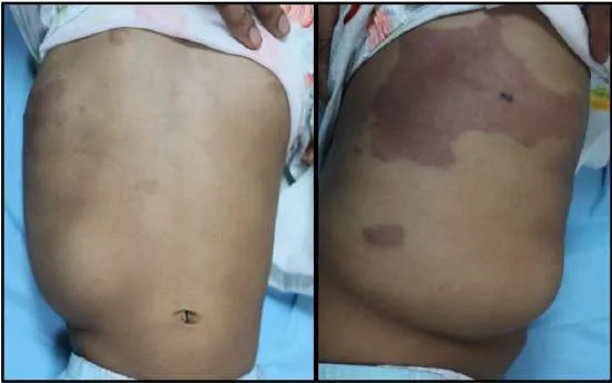
- Capillary–Venous Malformation (CVM) (Figure 9)
- Capillary–Lymphatic Malformation (CLM)
- Venous–Lymphatic Malformation (VLM)
- Capillary–Venous–Lymphatic Malformation (CVLM)
B. Combined Malformations with High-Flow Components
- Capillary–Arteriovenous Malformation (CM-AVM)
- Capillary–Venous–Arteriovenous Malformation
- Lymphatic–Venous–Arteriovenous Malformation
Syndromic Complex Vascular Malformations
Klippel–Trénaunay Syndrome (KTS)3,5,36: This syndrome is characterized by triad of capillary malformation, venous malformation, limb overgrowth. PIK3CA mutations are implicated in its pathogenesis and clinical features include limb hypertrophy, varicosities, pain, thrombosis, bleeding.
Parkes Weber Syndrome3,5,36: It is a combined vascular malformation with high-flow AV shunts caused by RASA1 mutations. Clinically, it presents as capillary malformation, AVFs, limb overgrowth, high-output cardiac failure.
CLOVES Syndrome3,5,36: It is characterized by Ccongenital lipomatous overgrowth, vascular malformations (CVLM), epidermal nevi, skeletal anomalies, caused be somatic PIK3CA mutations.
Proteus Syndrome3,5,36: It presents as sporadic overgrowth with mosaic distribution and the vascular features include capillary, venous, lymphatic malformations. Activating AKT1 mutation has been implicated as its cause.
MCAP Syndrome (Megalencephaly–Capillary Malformation)3,5,36: It’s features include diffuse capillary malformations, brain overgrowth, developmental delay, caused be PIK3CA mutations.
Diagnostic Evaluation: Diagnosis relies on clinical examination and imaging3,5,36: MRI is preferred to delineate extent and identify individual components. Doppler ultrasound differentiates low- and high-flow elements. Genetic testing is valuable in syndromic cases.3,5,36
Management Principles: Management involves multidisciplines tailored to lesion type and severity and may include Sclerotherapy, Embolization, Surgical debulking, Laser therapy and Targeted medical therapy (sirolimus, PI3K inhibitors in select cases).3,5,36
Prognosis: Prognosis varies widely depending on lesion extent and syndromic involvement.3,5,36 Some patients have stable disease, while others experience progressive complications requiring long-term care.3,5,36
REFERENCES
- Klement G, Fishman SJ. Vascular anomalies: hemangiomas and malformations. In: Coran AG, Adzick NS, Krummel TM, Laberge JM, Shamberger RC, Caldamone AA, editors. Pediatric Surgery. 7th ed. Elsevier/Mosby; 2012. p. 2094–2110.
- Virchow R. Die krankhaften Geschwülste. Vol 3. Berlin: August Hirschwald; 1863. p. 306–317.
- Mulliken JB, Glowacki J. Hemangiomas and vascular malformations in infants and children: a classification based on endothelial characteristics. Plast Reconstr Surg. 1982;69(3):412-422.
- Wassef M, Blei F, Adams D, et al. Vascular anomalies classification: recommendations from the ISSVA. Pediatrics. 2015;136(1):e203-e214.
- ISSVA Classification of Vascular Anomalies. International Society for the Study of Vascular Anomalies; updated 2018.
- Drolet BA, Trenor CC, Brandão LR, Chiu YE, Chun RH, Dasgupta R, et al. Consensus-derived practice standards for complicated Kaposiform hemangioendothelioma. Pediatrics. 2013;131(1):e263-e270.
- Dubois J, Garel L. Imaging and therapeutic approach of hemangiomas and vascular malformations in the pediatric age group. Pediatr Radiol. 1999;29(12):879-893.
- Dompmartin A, Vikkula M, Boon LM. Venous malformation: update on aetiopathogenesis, diagnosis and management. Phlebology. 2010;25(5):224-235.
- Legiehn GM, Heran MK. Venous malformations: classification, development, diagnosis, and interventional radiologic management. Radiol Clin North Am.
- 2008;46(3):545-597.
- Burrows PE, Laor T, Paltiel HJ, et al. Diagnostic imaging in the evaluation of vascular birthmarks. Dermatol Clin. 1998;16(3):45-488.
- Mulligan PR, Prajapati HJ, Martin LG, Patel TH. Vascular anomalies: classification, imaging characteristics and implications for interventional radiology treatment approaches. Br J Radiol. 2014;87(1035):20130392.
- Greene AK, Orbach DB. Management of arteriovenous malformations. Clin Plast Surg. 2011;38(1):95-106.
- Cahill AM, Nijs EL. Pediatric vascular anomalies: an update. Semin Intervent Radiol. 2013;30(3):215-223.
- Dubois J, Alison M. Vascular anomalies: what a radiologist needs to know. Pediatr Radiol. 2010;40(6):895-905.
- Léauté-Labrèze C, Dumas de la Roque E, Hubiche T, Boralevi F, Thambo JB, Taïeb A. Propranolol for severe hemangiomas of infancy. N Engl J Med. 2008;358(24):2649–
- doi:10.1056/NEJMc0708819
- Drolet BA, Esterly NB, Frieden IJ. Hemangiomas in children. N Engl J Med.
- 1999;341(3):173–81. doi:10.1056/NEJM199907153410307
- Frieden IJ, Haggstrom AN, Drolet BA, Mancini AJ, Friedlander SF, Marchant GE, et al. Infantile hemangiomas: current knowledge, future directions. Pediatrics.
- 2005;115(4):883–97. doi:10.1542/peds.2004-2346
- Krowchuk DP, Frieden IJ, Mancini AJ, Darrow DH, Blei F, Greene AK, et al. Clinical practice guideline for the management of infantile hemangiomas. Pediatrics.
- 2019;143(1):e20183475. doi:10.1542/peds.2018-3475.
- Hoeger PH, Harper JI, Baselga E, Bonnet D, Guibaud L, Mazereeuw-Hautier J, et al. Treatment of infantile haemangiomas: recommendations of a European expert group. Eur J Pediatr. 2015;174(7):85–65. doi:10.1007/s00431-015-2570-0
- Bauman NM, McCarter RJ, Guzzetta PC, Perkins JA, Burrows PE, Berenstein A, et al. Propranolol vs prednisolone for symptomatic proliferating infantile hemangiomas. Otolaryngol Head Neck Surg. 2014;150(5):789–95. doi:10.1177/0194599814521926
- Boon LM, Enjolras O, Mulliken JB. Congenital hemangioma: evidence of accelerated involution. J Pediatr. 1996;128(3):329–35. doi:10.1016/S0022-3476(96)70398-1
- Mulliken JB, Enjolras O. Congenital hemangiomas and infantile hemangioma: missing links. J Am Acad Dermatol. 2004;50(6):875–82. doi:10.1016/j.jaad.2003.12.017
- Enjolras O, Mulliken JB, Boon LM, Vikkula M. Noninvoluting congenital hemangioma: a rare cutaneous vascular anomaly. Plast Reconstr Surg.
- 2001;107(7):1647–54. doi:10.1097/00006534-200106000-00010
- North PE, Waner M, Mizeracki A, Mihm MC Jr. GLUT-1: a newly discovered immunohistochemical marker for juvenile hemangiomas. Hum Pathol. 2000;31(1):11–
- doi:10.1016/S0046-8177(00)80199-6
- Baselga E, Cordisco MR, Garzon MC, Marcuzzi D, Edgerton MT, Mullick FG, et al. Rapidly involuting congenital hemangioma and partially involuting congenital hemangioma: clinical and histopathologic features. J Am Acad Dermatol.
- 2008;58(5):757–62. doi:10.1016/j.jaad.2007.12.026
- Christison-Lagay ER, Fishman SJ. Hepatic hemangiomas. Semin Pediatr Surg. 2014;23(4):194–202. doi:10.1053/j.sempedsurg.2014.06.008
- Chung EM, Cube R, Lewis RB, Conran RM. From the archives of the AFIP: pediatric liver masses—radiologic-pathologic correlation part 1. Benign tumors. Radiographics. 2010;30(3):801–826. doi:10.1148/rg.303095173
- Haggstrom AN, Drolet BA, Baselga E, Chamlin SL, Garzon MC, Horii KA, et al. Prospective study of infantile hemangiomas: demographic, prenatal, and perinatal characteristics. J Pediatr. 2007;150(3):291–294. doi:10.1016/j.jpeds.2006.12.003
- North PE, Waner M, James CA, Mizeracki A, Mihm MC Jr. Congenital nonprogressive hemangioma: a distinct clinicopathologic entity unlike infantile hemangioma. Arch Dermatol. 2001;137(12):1607–1620. doi:10.1001/archderm.137.12.1607
- Kassarjian A, Fishman SJ, Fox VL, Bowden DW, Graf JL, Filston HC. Hepatic hemangiomas in infancy: clinical and imaging findings. AJR Am J Roentgenol. 2004;182(4):1003–1013. doi:10.2214/ajr.182.4.1821003
- Huang SA, Tu HM, Harney JW, Venihaki M, Liu F, Butte AJ, et al. Severe hypothyroidism caused by type 3 iodothyronine deiodinase in infantile hepatic hemangiomas. N Engl J Med . 2000;343(3):185 –189. doi:10.1056/NEJM200007203430304
- Nelson JC, Clark SJ, Borut DL, Tomei RT, Carlton EI. Consumptive hypothyroidism associated with hepatic hemangiomas. N Engl J Med. 1987;317(11):665–668. doi:10.1056/NEJM198709103171105
- Brisse HJ, Orbach D, Savignoni A, Launay EA, Chaumoitre K, Dhelalf F, et al. Imaging of hepatic hemangiomas in infants. Pediatr Radiol. 2012;42(5):613–624. doi:10.1007/s00247-011-2321-4
- Léauté-Labrèze C, Hoeger P, Mazereeuw-Hautier J, Guibaud L, Baselga E, Posiunas G, et al. A randomized, controlled trial of oral propranolol in infantile hemangioma. N Engl J Med. 2015;372(8):735–746. doi:10.1056/NEJMoa1404710
- Weiss SW, Goldblum JR. Enzinger and Weiss’s Soft Tissue Tumors. 6th ed. Philadelphia: Elsevier; 2014.
- Enjolras O, Wassef M, Chapot R. Colour Atlas of Vascular Tumours and Vascular Malformations. Cambridge: Cambridge University Press; 2007.
- Croteau SE, Liang MG, Kozakewich HP, Perez-Atayde AR, Alomari AI, Fishman SJ, et al. Kaposiform hemangioendothelioma: atypical features and clinical outcomes in a cohort of 107 patients. J Pediatr. 2013;162(1):142–7. doi:10.1016/j.jpeds.2012.06.044
- Fletcher CDM, Bridge JA, Hogendoorn PCW, Mertens F, editors. WHO Classification of Tumours of Soft Tissue and Bone. 5th ed. Lyon: IARC; 2020.
- O’Rafferty C, O’Connor R, McKenna P, Fitzgerald M, Kieran M, Ryan K, et al. Kasabach–Merritt phenomenon: pathophysiology and management. Br J Haematol. 2015;169(2):173–84. doi:10.1111/bjh.13250
- Ji Y, Chen S, Yang K, Xia C, Li L, Lin X, et al. Sirolimus for the treatment of kaposiform hemangioendothelioma and tufted angioma: long-term outcomes. J Am Acad Dermatol. 2020;83(2):540–548. doi:10.1016/j.jaad.2019.12.046.
- Triana P, Dore M, Cerezo VN, Manuel Cervantes, Alejandra Vilanova Sánchez, Miriam Miguel Ferrero, et al. Sirolimus in the treatment of vascular anomalies. Eur J Pediatr Surg. 2017;27(1):86–90. doi:10.105/s-0036-1581115
- Adams DM, Trenor CC, Hammill AM, Vinks AA, Patel MN, Chaudry G,et al. Efficacy and safety of sirolimus in the treatment of complicated vascular anomalies. Pediatrics. 2016;137(2):e20153257. doi:10.1542/peds.2015-3257
- Venot Q, Blanc T, Rabia SH, Berteloot L, Ladraa S, Duong JP,et al. Targeted therapy in patients with PIK3CA-related overgrowth syndrome. Nature. 2018;58(7711):540–
- doi:10.1038/s41586-018-0217-9
- Canaud G, Guitierrez JCL, Irvine AD, Vabres P, Hansford JR, Ankrah N, et al. Alpelisib for treatment of severe PIK3CA-related vascular malformations. N Engl J Med. 2021;384(17):1585–95. doi:10.1056/NEJMoa2021326
- Greene AK, Goss JA. Vascular anomalies: from a clinicohistologic to a genetic
| framework. Plast Reconstr Surg. 2018;141(5):709e– doi:10.1097/PRS.0000000000004321 | 717e. | |
|---|---|---|
| 46. | Mills SE, Cooper PH, Fechner RE. Lobular capillary hemangioma: the underlying lesion of pyogenic granuloma. Am J Surg Pathol. 1980;4(5):470 doi:10.1097/00000478-198010000-00008 | –9. |
| 47. | Patrice SJ, Wiss K, Mulliken JB. Pyogenic granuloma (lobular capillary hemangioma): a clinicopathologic study of 178 cases. Pediatr Dermatol. 1991;8(4):267 doi:10.1111/j.1525-1470.1991.tb00920.x | –76. |
48. Jafarzadeh H, Sanatkhani M, Mohtasham N. Oral pyogenic granuloma: a review. J Oral Sci. 2006;48(4):167–75. doi:10.2334/josnusd.48.167
49. Mulliken JB, Young AE. Vascular Birthmarks: Hemangiomas and Malformations. Philadelphia: WB Saunders; 1988;320.
50. Greene AK, Goss JA. Vascular anomalies: from a clinicohistologic to a genetic framework. Plast Reconstr Surg. 2018;141(5):709e –717e. doi:10.1097/PRS.0000000000004321
51. Shirley MD, Tang H, Gallione CJ, Baugher JD, Frelin LP, Cohen B, North PE, Marchuk DA, Comi AM, Pevsner J. Sturge–Weber syndrome and port-wine stains caused by somatic mutation in GNAQ. New England Journal of Medicine. 2013 May 23;368(21):1971-9.